Pinworms: Key Facts at a Glance
Pinworms are tiny, white, highly contagious parasitic roundworms that live in the human gut. The hallmark symptom is intense itching around the anus at night, which can disturb sleep. They are highly treatable, generally not dangerous, and do not reflect poor personal hygiene.
- How they spread: Eggs lodge under the fingernails when a child scratches and are then swallowed. They also spread through bed sheets, pajamas, underwear, towels, and shared surfaces, where they can survive for two to three weeks.
- Other signs in children: Irritability and tiredness during the day, teeth grinding during sleep, and bedwetting in a child who was already toilet trained.
- Diagnosis: The tape test, done immediately on waking and before bathing or using the toilet. Repeating it on three consecutive mornings raises accuracy to over ninety percent.
- Treatment: Mebendazole, albendazole, or pyrantel pamoate. Two doses are mandatory, with the second given exactly two weeks after the first, because these medicines do not kill the eggs.
- Treat the whole household: If one person is diagnosed, every person living in the household must be treated at the same time, or the infection keeps returning.
- Daily hygiene: Shower on waking instead of taking a bath, clip fingernails short, and wash bedding, pajamas, and underwear in the hottest water available without shaking them first.
- Habits to stop: Discourage nail biting and thumb sucking until the eradication process is complete.
- A Comprehensive Medical Guide to Understanding Pinworms
- What Exactly Are Pinworms?
- The Intricate Life Cycle of Pinworms
- Primary Modes of Transmission for Pinworms
- Diagnostic Procedures and Medical Evaluation of Pinworms
- Evidence-Based Treatment Strategies
- Rare but Serious Complications Associated with Pinworms
- Frequently Asked Questions
- Scientific Sources
A Comprehensive Medical Guide to Understanding Pinworms
Encountering a parasitic infection within your household can be an incredibly distressing experience for any parent, caregiver, or individual. Among the most prevalent of these parasitic challenges are Pinworms, a type of intestinal nematode that has coexisted with human beings throughout our evolutionary history. Although the discovery of these organisms can cause significant psychological distress and anxiety, it is essential to understand that they are highly treatable and do not reflect poor personal hygiene or a lack of cleanliness. This comprehensive guide aims to demystify the biological nature of these organisms, explore their intricate life cycle, detail the clinical manifestations they provoke, and provide evidence-based strategies for eradication and prevention. By equipping yourself with scientifically backed information, you can approach this common childhood ailment with confidence, ensuring the health, comfort, and well-being of your entire family.
What Exactly Are Pinworms?
To effectively manage and eradicate any medical condition, one must first deeply understand the biological entity responsible for it. Pinworms are small, white, thread-like parasitic roundworms that belong to the nematode family Oxyuridae. Scientifically classified and referred to as Enterobius vermicularis, these organisms are strictly human pathogens. This means that humans are their only natural host; you cannot catch this specific parasite from your household pets, nor can you transmit it to them. The historical relationship between humans and this specific parasite is extensive and fascinating. Archaeological evidence, including fossilized human coprolites and ancient mummies, indicates that these organisms have been infecting human digestive tracts for many thousands of years.
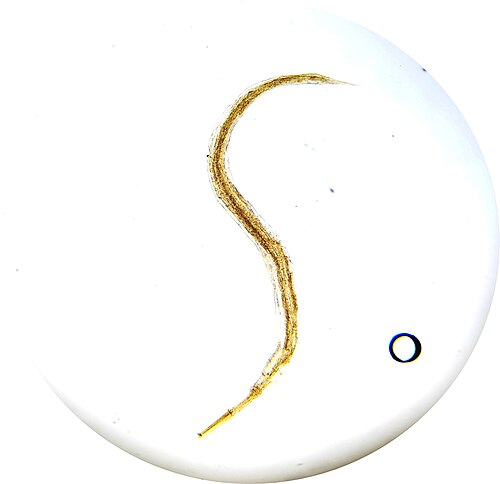
The physical appearance of these parasites is quite distinct and serves as the primary basis for visual diagnosis. The adult female worms are notably larger than their male counterparts, typically measuring between eight and thirteen millimeters in length and approximately half a millimeter in thickness. They possess a characteristic sharply pointed, pin-like posterior end, which is the anatomical feature that earned them their common name. The adult males are considerably smaller, usually measuring only two to five millimeters in length, and feature a distinctly curved tail end.
Recognizing pinworms parasites and Their Anatomical Characteristics
When analyzing pinworms parasites under a laboratory microscope, medical professionals look for specific anatomical markers. These include cephalic alae, which are prominent ridge-like structures located at the head region of the worm, and a muscular esophagus with a distinct posterior bulb. The eggs produced by these parasites are microscopic, measuring roughly fifty to sixty micrometers by twenty to thirty micrometers. They are translucent and possess a highly specialized, sticky outer shell that is flattened on one side, giving them a unique asymmetrical appearance often compared to a slice of bread. This sticky outer membrane is a crucial evolutionary adaptation, allowing the eggs to adhere firmly to the human skin, clothing, and environmental surfaces, thereby facilitating rapid transmission.
The Intricate Life Cycle of Pinworms
The life cycle of Pinworms is a fascinating, albeit unsettling, biological process that takes place entirely within the human gastrointestinal tract. Unlike many other complex parasites that require intermediate hosts such as snails, pigs, or mosquitoes to complete their developmental stages, this organism relies solely on a single human host. The entire life cycle, spanning from the ingestion of an infective egg to the death of the adult worm, generally takes about four to eight weeks to complete.
How a pinworm infection Progresses in the Human Digestive System
The cycle of a pinworm infection officially begins when a human host accidentally ingests embryonated, infective eggs. Once these microscopic eggs are swallowed, they travel down the esophagus and safely pass through the harsh, acidic environment of the stomach. Upon reaching the duodenum—the uppermost section of the small intestine—the digestive fluids soften the sticky outer membrane of the eggs, prompting them to hatch.
The newly emerged larvae are incredibly small, measuring merely one hundred and forty to one hundred and fifty micrometers. These highly active larvae begin a slow migration down the length of the small intestine, progressing toward the colon. During this extensive journey, the larvae undergo multiple molting phases, shedding their outer cuticles as they rapidly mature into adult male and female worms. Once they reach the ileocecal region—the anatomical junction where the small intestine meets the large intestine, encompassing the cecum and the appendix—the adult worms settle down to mate.
Following reproduction, the male worms typically die and are harmlessly passed out of the body within the fecal matter. The gravid (pregnant) females, however, attach themselves to the mucosal lining of the colon, where they absorb nutrients and allow the thousands of eggs within their bodies to mature. A single gravid female can carry an astonishing payload of ten thousand to sixteen thousand eggs. When the eggs are fully developed, the female detaches from the intestinal wall and begins a long migration down the descending colon and rectum.
This migration generally occurs late at night or during the early hours of the morning when the human host is asleep and resting quietly. The female worm slowly emerges from the anal sphincter and enters the oxygen-rich environment of the perianal folds. Here, she begins to forcefully expel her eggs onto the surrounding skin. The physical act of laying eggs, combined with the irritating, glue-like substance secreted by the female to adhere the eggs to the skin, triggers an intense localized inflammatory and allergic response. After completely emptying her reproductive tract, the female worm becomes opaque, quickly dies, and disintegrates. Within just four to six hours of being deposited in the warm, humid environment of the perianal region, the eggs mature and become fully infective, ready to begin the cycle anew.
Global Epidemiology and Prevalence of Pinworms
When discussing the epidemiology of Pinworms, it is crucial to recognize that this is not a localized or regional health issue, but rather a massive global phenomenon. Medical estimates suggest that more than one billion people worldwide are currently infected with this parasite. Unlike many other soil-transmitted helminths and parasitic infections that are strictly confined to developing nations with poor sanitation infrastructure, this specific nematode thrives in both developing and highly developed, affluent countries. It is recognized as the most common parasitic worm infection in the United States, Western Europe, and Oceania.
The Spread of pinworms enterobiasis Across Different Climates
The widespread transmission of pinworms enterobiasis is remarkably uniform across various geographical climates, though it tends to peak in areas with moderate temperatures. The highest prevalence rates are consistently observed in preschool and school-aged children, typically between the ages of four and eleven years old. Studies have shown that up to fifty percent of children in certain European and North American schooling systems may harbor the parasite at any given time. The infection rates are inherently linked to the behavioral habits of young children, such as frequent finger-sucking, inadequate hand hygiene, and a lack of understanding regarding personal boundaries and sanitary practices. Furthermore, because the eggs are highly resilient in indoor environments, the infection frequently sweeps through entire family units, daycares, kindergartens, and institutionalized care facilities.
Primary Modes of Transmission for Pinworms
Understanding exactly how Pinworms are transmitted from one individual to another is the absolute cornerstone of both preventing initial outbreaks and stopping frustrating recurrent infections. The transmission is highly efficient and occurs primarily through the fecal-oral route. This means that the microscopic eggs must be transferred from the perianal region of an infected individual to the mouth of the same person or a new host.
Identifying Common Pinworms causes in Children and Adults
One of the most frequent Pinworms causes is the act of autoinfection. When the female worm lays her eggs at night, the host experiences intense pruritus (itching). In response, the host—often a sleeping child—will instinctively scratch the perianal area. During this vigorous scratching, the highly adhesive eggs become firmly lodged beneath the fingernails and onto the fingertips. If the child subsequently bites their nails, sucks their thumb, or handles food without thoroughly washing their hands with warm water and soap, they swallow the eggs, thereby instantly re-infecting themselves and guaranteeing the continuation of the parasite’s life cycle.
Beyond direct autoinfection, indirect transmission via fomites (inanimate objects) is incredibly common. The microscopic eggs can easily detach from the perianal skin and heavily contaminate the host’s immediate environment. Bed sheets, pajamas, underwear, and towels become primary reservoirs for the eggs. From there, the eggs can be transferred to shared household items, including bathroom faucets, toilet seats, doorknobs, and children’s toys. The eggs are remarkably resilient and can survive in a cool, humid indoor environment for two to three weeks, patiently waiting to be picked up by the hands of a new, unsuspecting host. Furthermore, while relatively rare, airborne transmission is a documented possibility. Because the eggs are exceptionally light and microscopic, vigorous activities such as shaking out contaminated bed linens, vigorously dusting a heavily infested room, or sweeping can launch the eggs into the air. Once airborne, these eggs can be inhaled through the nose or mouth and subsequently swallowed, leading to a new infection without any direct physical contact ever occurring. Finally, a unique transmission phenomenon known as retroinfection can occur. In this scenario, the eggs hatch on the perianal skin itself, and the newly emerged larvae crawl back through the anal sphincter into the rectum and colon to mature, bypassing the oral ingestion route entirely.
Recognizing the Clinical Signs of Pinworms
The clinical presentation of Pinworms can range from completely asymptomatic to severely distressing. It is a well-documented medical fact that approximately one-third to forty percent of all infected individuals carry the parasite without ever displaying a single noticeable symptom. These asymptomatic carriers are particularly concerning from a public health perspective, as they act as silent reservoirs, unknowingly shedding microscopic eggs into their environment and facilitating the continuous spread of the parasite to more vulnerable individuals.
Primary Pinworms symptoms to Watch For in Your Household
For those who do exhibit clinical signs, the hallmark of Pinworms symptoms is an intense, agonizing itchiness in the perianal or perineal region, scientifically termed pruritus ani. This localized itching is almost exclusively nocturnal, corresponding perfectly with the biological habits of the gravid female worm migrating to the exterior folds of the anus to deposit her eggs. The severity of this itching should never be underestimated; it can cause profound discomfort and lead to significant sleep disturbances.
Children suffering from this nocturnal irritation frequently exhibit secondary behavioral and physical symptoms as a direct result of sleep deprivation. Parents may notice their child becoming increasingly irritable, restless, and fatigued during the daytime. Furthermore, the physical discomfort can lead to bruxism (the unconscious grinding of teeth during sleep) and unexpected episodes of nocturnal enuresis (bedwetting) in children who had previously been successfully toilet trained.
Detailed pinworm disease symptoms and pinworm infection symptoms
When examining detailed pinworm disease symptoms, medical professionals also look out for the dermatological consequences of chronic scratching. Continuous, aggressive scratching of the sensitive perianal skin invariably leads to excoriations (deep scratch marks) and the breakdown of the skin’s natural protective barrier. This creates an ideal entry point for opportunistic skin bacteria, frequently resulting in painful, inflamed secondary bacterial infections, perianal folliculitis, or even the formation of ischiorectal abscesses. While less common, some patients report experiencing vague, intermittent gastrointestinal complaints. These pinworm infection symptoms can occasionally manifest as mild abdominal pain, generalized nausea, a sudden loss of appetite, or unexplainable weight loss, though these are typically only observed in cases involving an exceptionally heavy parasitic burden.
Diagnostic Procedures and Medical Evaluation of Pinworms
Diagnosing an active infestation of Pinworms requires a specific approach that differs significantly from the diagnostic protocols used for other common intestinal parasites. Because these specific nematodes do not routinely lay their eggs within the intestinal lumen, relying on standard stool sample analyses is famously ineffective and will frequently yield false-negative results.
Spotting pinworms in poop and Identifying a pinworm in stool
Many anxious parents meticulously examine their children’s diapers or toilet bowls, attempting to spot pinworms in poop. While it is occasionally possible to see the adult worms crawling on the absolute outer surface of a freshly passed bowel movement, finding a pinworm in stool is generally considered a rare and unreliable diagnostic occurrence. The adult worms tightly anchor themselves to the mucosal lining of the cecum and are generally only expelled in the feces when the host is experiencing an unusually massive, overwhelming parasitic burden.
What Does feces with pinworms Actually Look Like?
If you do happen to observe feces with pinworms, the parasites will appear as tiny, pale, squirming threads, roughly the size of a standard staple, moving erratically across the surface of the stool. However, because this visual confirmation is rare, the gold standard for medical diagnosis is the cellophane tape test.
This highly effective diagnostic procedure is best performed immediately upon the patient waking up in the morning, crucially before they have bathed, showered, or used the toilet, as these activities will wash away the diagnostic evidence. A piece of clear, transparent adhesive tape is firmly pressed, sticky side down, against the perianal skin folds. The tape is then carefully removed and applied flat to a sterile glass laboratory slide. A trained medical technician or physician will then examine this slide under a high-powered microscope. If the characteristic asymmetrical, slice-of-bread-shaped eggs are visualized, the diagnosis is definitively confirmed. Because the female worms do not migrate and lay eggs every single night, a single tape test only has a sensitivity of about fifty percent. Therefore, medical guidelines strongly recommend repeating the tape test on three consecutive mornings, which dramatically increases the diagnostic accuracy to over ninety percent.
Differential Diagnosis
When a patient presents with severe perianal itching, healthcare providers must consider a comprehensive differential diagnosis to ensure accurate treatment. Conditions such as atopic dermatitis, psoriasis, fungal infections (like candidiasis), hemorrhoids, and even poor wiping hygiene can mimic the intense pruritus caused by these parasites. Therefore, microscopic confirmation is always the preferred route before initiating pharmacological therapy.
Clarifying the Debate: Threadworms vs pinworms
There is often significant linguistic confusion in the medical literature regarding the terminology of Threadworms vs pinworms. It is essential to clarify that in the United Kingdom, Australia, and New Zealand, the term “threadworm” is the standard colloquial name used by the general public and the National Health Service (NHS) to describe Enterobius vermicularis. Therefore, in these specific geographic regions, the two terms are entirely synonymous and refer to the exact same parasite.
However, in the United States and the broader field of international parasitology, the term “threadworm” is strictly reserved for a completely different and vastly more dangerous species of nematode known as Strongyloides stercoralis. Strongyloides can cause severe, life-threatening systemic infections, particularly in immunocompromised individuals. Therefore, relying on accurate scientific nomenclature (Enterobius vermicularis) is vital for clear medical communication and ensuring the administration of the correct therapeutic protocols.
Evidence-Based Treatment Strategies
Fortunately, the eradication of Pinworms is highly achievable using modern pharmacological interventions. The primary goal of medical therapy is to completely eliminate the adult worms from the gastrointestinal tract, thereby halting the nocturnal deposition of eggs and breaking the transmission cycle.
Protocols for pinworm treatment and Comprehensive pinworms treatment
The most universally prescribed medications for pinworm treatment include Mebendazole, Albendazole, and Pyrantel Pamoate. These anthelmintic (anti-worm) drugs are exceptionally effective, generally boasting cure rates exceeding ninety to ninety-five percent when administered correctly.
Mebendazole and Albendazole work by directly attacking the cellular infrastructure of the parasite. They specifically inhibit the polymerization of a protein called tubulin within the worm’s cells. Without functioning tubulin, the parasite loses its ability to uptake glucose, quickly depleting its energy reserves, which inevitably leads to paralysis and death. Pyrantel Pamoate, which is widely available as an over-the-counter pinworms treatment in many countries, operates via a different mechanism. It acts as a depolarizing neuromuscular blocking agent, causing sudden, rigid, and irreversible paralysis of the adult worms. Once paralyzed, the worms lose their grip on the intestinal wall and are naturally flushed out of the host’s body during normal bowel movements.
Regardless of which specific medication is chosen, a strict two-dose regimen is absolutely mandatory for clinical success. The initial dose of the medication is highly effective at killing all of the adult worms currently residing in the colon. However, none of these medications are ovicidal, meaning they cannot penetrate the thick shell of the eggs, nor can they effectively kill the immature larvae that are still developing within the upper digestive tract. Therefore, a mandatory second dose must be administered exactly two weeks after the initial dose. This highly strategic timing ensures that any eggs that survived the first round of treatment have hatched and matured into vulnerable adult worms, allowing the second dose of medication to completely wipe them out before they are old enough to begin reproducing.
“Clinical failure in parasite eradication is rarely due to drug resistance; rather, it is almost entirely attributed to the failure to administer the critical second dose at the two-week mark, or failing to treat asymptomatic household members.” — Standard Pediatric Infectious Disease Guidelines.
Environmental Control and Eradicating Pinworms from Your Home
While pharmacological intervention is incredibly effective at clearing the parasite from the human body, medicine alone cannot prevent immediate reinfection if the patient’s physical environment remains heavily contaminated with microscopic eggs. Therefore, treating Pinworms requires a rigorous, multi-faceted approach that combines medication with strict environmental hygiene and behavioral modifications.
Understanding the importance of a single pinworm eradication
The most crucial rule of environmental management is that if one single person in a household is diagnosed with this infection, every single person living in that household must be treated simultaneously. Because the transmission rate is so incredibly high, and asymptomatic carriage is so common, attempting to selectively treat only the scratching child will inevitably lead to a cycle of endless reinfection known as “ping-ponging” within the family unit.
To systematically cleanse the home environment, families must implement a comprehensive daily hygiene checklist.
| Daily Hygiene Checklist for Parasite Eradication | Action Required | Frequency |
| Morning Bathing | Shower immediately upon waking. Do not take a bath, as eggs can float in bathwater and re-adhere to the skin. | Daily, for at least two weeks. |
| Nail Care | Clip all fingernails extremely short. Scrub vigorously under the nails with a stiff brush and antibacterial soap. | Daily monitoring. |
| Laundry Protocols | Carefully roll up (do not shake) bedsheets, pajamas, and underwear. Wash them in the hottest water setting available and dry on high heat. | Daily, especially during the first week of treatment. |
| Surface Disinfection | Wipe down toilet seats, flush handles, bathroom faucets, and doorknobs with hot water and strong disinfectants. | Daily. |
| Environmental Control | Open bedroom curtains and blinds during the day. The parasite eggs are highly sensitive to natural sunlight and ultraviolet radiation. | Daily. |
| Vacuuming | Thoroughly vacuum all carpets, rugs, and soft furnishings to remove eggs that have fallen to the floor. Use a damp cloth for dusting. | Every 2-3 days. |
It is absolutely critical to avoid shaking out bed linens or pajamas before washing them. Shaking contaminated fabrics acts like a dispersal mechanism, launching the invisible eggs into the air where they can settle on clean surfaces or be accidentally inhaled. Furthermore, parents must actively discourage children from biting their nails, sucking their thumbs, or engaging in any hand-to-mouth behaviors until the eradication process is fully complete.
Rare but Serious Complications Associated with Pinworms
While a diagnosis of Pinworms is generally considered more of a severe nuisance and a psychological burden than a dangerous medical emergency, exceptional cases involving aberrant parasitic migration can lead to significant and concerning health complications.
Because the female worms blindly crawl outward from the anus at night, they can occasionally lose their way and migrate into adjacent anatomical orifices. In young girls and adult women, the female worms frequently migrate forward into the vulva and the vagina. This ectopic migration introduces intestinal bacteria into the sterile reproductive tract, leading to intense vulvovaginitis, characterized by severe vaginal itching, abnormal discharge, and localized pain. If left untreated, the worms can theoretically travel further up the reproductive tract, penetrating the uterus and the fallopian tubes, potentially triggering pelvic inflammatory disease (PID) or localized granulomas.
Another well-documented, albeit controversial, complication is the relationship between this parasite and acute appendicitis. Because the cecum and the appendix are the primary anatomical habitats for the adult worms, a massive, overwhelming parasitic burden can physically block the narrow lumen of the appendix. This mechanical obstruction can lead to appendiceal inflammation, perfectly mimicking the clinical presentation of acute appendicitis. Pathological examinations of surgically removed appendices occasionally reveal the presence of adult worms nested deep within the inflamed tissue. Furthermore, chronic parasitic infections have been linked to instances of eosinophilic colitis, a condition characterized by inflammation of the large intestine resulting from an abnormal accumulation of specific white blood cells attempting to fight off the continuous parasitic presence.
Frequently Asked Questions
How do you get pinworms?
You acquire this parasitic infection by accidentally swallowing or inhaling their microscopic eggs. This usually happens when an infected person scratches their itchy perianal area, trapping the highly adhesive eggs under their fingernails, and subsequently touches shared surfaces, food, or another person. If you touch those contaminated objects—such as doorknobs, toys, or bed linens—and then unconsciously touch your mouth or eat without washing your hands, the eggs enter your digestive system, leading to a full-blown infection.
What are pinworms?
They are tiny, white, highly contagious parasitic roundworms that exclusively infect the human gastrointestinal tract. Scientifically classified as Enterobius vermicularis, the adult females grow to about the size of a standard staple and migrate to the perianal area at night to lay thousands of microscopic eggs. These parasites are universally common, especially among young school-aged children, and cause intense nocturnal itching, though they rarely lead to severe or life-threatening medical complications.
How to get rid of pinworms?
Eradicating these parasites requires a strict, dual approach combining pharmacological treatment and rigorous household hygiene. You must take an anti-parasitic medication (like Mebendazole or Pyrantel Pamoate) in two distinct doses spaced exactly two weeks apart to kill both current adult worms and those that hatch later. Furthermore, you must simultaneously treat every member of your household, wash all bedding and underwear in extremely hot water daily, mandate morning showers, and strictly enforce short fingernails and frequent handwashing.
Can pinworms kill you?
No, it is highly improbable for this specific parasite to cause a fatal outcome in a human host. While they are incredibly annoying, cause immense psychological distress, and lead to severe sleep deprivation due to intense itching, they are fundamentally non-invasive organisms that remain confined to the digestive tract. In extremely rare, atypical cases, they can cause complications like appendicitis or pelvic inflammatory disease by wandering into the wrong organs, but even these scenarios are almost never life-threatening when managed with standard modern medical care.
What do pinworms look like?
To the naked eye, the adult parasites look like very small, thin, white pieces of thread or slightly wriggling staples, measuring roughly eight to thirteen millimeters in length. If you inspect the perianal area of an infected child at night using a flashlight, you may see them actively moving on the surface of the skin. Their eggs, however, are completely invisible without a laboratory microscope and are only detectable using specific medical procedures like the cellophane tape test.
Are pinworms dangerous?
Generally speaking, they are not considered medically dangerous; they are classified by healthcare professionals as a benign, highly treatable nuisance condition. The primary harm they cause is intense physical discomfort, skin excoriation from chronic scratching, and the resulting fatigue and irritability stemming from interrupted sleep patterns. While secondary bacterial skin infections or rare vaginal migrations require medical attention, the parasites themselves do not cause systemic disease, organ failure, or permanent internal damage.
The following posts may interest you
Cryptosporidiosis: Symptoms, Causes, and Treatment Guide
Giardiasis: A Complete Guide to Symptoms and Causes
Understanding Diarrhea: Causes, Symptoms, and Effective Treatments
Disclaimer: The content provided on BabyKidsHealth.com is for informational purposes only and is not intended as a substitute for professional medical advice, diagnosis, or treatment. Always seek the advice of your physician or other qualified health provider with any questions you may have regarding a medical condition.
Sources
About Pinworm Infection (Centers for Disease Control and Prevention, CDC)
https://www.cdc.gov/pinworm/about/index.html
Pinworms (American Academy of Pediatrics, HealthyChildren.org)
https://www.healthychildren.org/English/health-issues/conditions/abdominal/Pages/pinworms.aspx
The Diagnosis and Treatment of Pinworm Infection
https://pmc.ncbi.nlm.nih.gov/articles/PMC6522669
Pinworm Infestation in Children: The Problem and Its Treatment
https://pmc.ncbi.nlm.nih.gov/articles/PMC1848787
Knowledge and Practice of Pinworm Infection in Preschool Children, Jiangsu Province, China, 2019–2020
https://ajph.aphapublications.org/doi/full/10.2105/AJPH.2022.307067
Manifestations of pinworms
https://pmc.ncbi.nlm.nih.gov/articles/PMC1930483
Pin Worms Presenting as Suspected Crohn’s Disease
https://pmc.ncbi.nlm.nih.gov/articles/PMC4612463
Prevalence of Enterobius vermicularis Infection among Preschool Children in Kindergartens of Taipei City, Taiwan in 2008
https://pmc.ncbi.nlm.nih.gov/articles/PMC2688803
Enterobius vermicularis and allergic conditions in Norwegian children
Transmission of Dientamoeba fragilis: pinworm or cysts?
https://www.cell.com/trends/parasitology/abstract/S1471-4922(14)00006-3
Pinworm infections associated with risk of psychiatric disorders-A nationwide cohort study in Taiwan: Pinworm infections and psychiatric disorders
https://www.sciencedirect.com/science/article/pii/S0010440X19300100