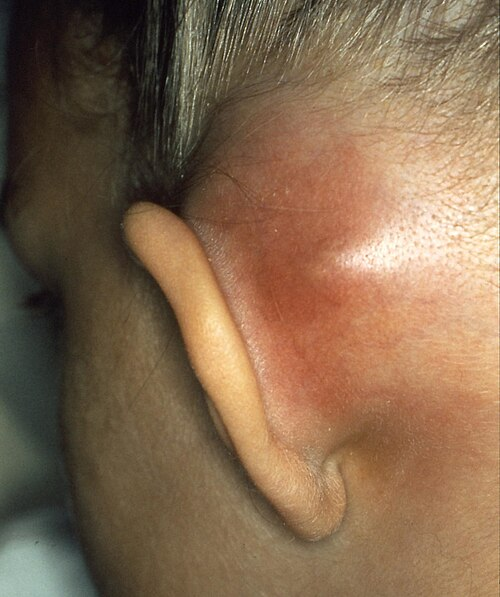
Mastoiditis: Key Facts at a Glance
Mastoiditis is a serious bacterial infection of the mastoid air cells, the honeycomb-like bony spaces in the temporal bone directly behind the ear. It most commonly develops when a middle ear infection goes untreated or only partially responds to therapy, allowing bacteria to spread into the adjacent bone. It is uncommon today, but it remains an emergency that needs prompt hospital care.
- Main symptoms: Persistent ear pain, high fever, headache, ear drainage, and redness, swelling, and tenderness over the bone behind the ear. As the swelling grows, it can push the ear forward and outward.
- Signs in babies and toddlers: Symptoms may be subtle: irritability, poor feeding, trouble sleeping, high fever, and repeated pulling or swatting at the affected side of the head. Recent antibiotic use can hide the fever.
- Who is most at risk: Infants and children under two years of age, with peak incidence around twelve months. Recurrent ear infections, a weakened immune system, and an underlying cholesteatoma (an abnormal skin growth in the middle ear) also raise the risk.
- Diagnosis: An ear examination, blood tests that measure inflammation, and a high-resolution CT scan of the temporal bone, which shows whether the bone is breaking down. An MRI is added when spread inside the skull is suspected.
- Treatment: Hospital admission for intravenous antibiotics, usually with a small drainage procedure on the eardrum (myringotomy with a tympanostomy tube). If there is no improvement within 48 hours, or CT shows bone destruction or an abscess, surgery to remove infected bone (cortical mastoidectomy) is performed. Oral antibiotics alone are not enough at the start.
- Emergency warning signs: High spiking fevers with shaking chills, increasing drowsiness, vision changes, confusion, facial weakness or asymmetry, abnormal eye movements, or a stiff neck can mean the infection is spreading and need immediate emergency care.
- Prevention: Keep routine childhood vaccinations up to date, including pneumococcal vaccines. Seek medical care when ear pain or fever does not improve within 48 to 72 hours, finish prescribed antibiotics as directed, and keep children away from tobacco smoke.
Comprehensive Clinical Guide to Mastoiditis: Etiology, Diagnosis, and Management
Mastoiditis is an acute or chronic suppurative bacterial infection that targets the mastoid air cells within the temporal bone of the human skull. This serious clinical entity develops when an unresolved middle ear infection extends beyond the mucosal confines of the tympanic cavity, directly invading the porous, honeycomb-like osseous compartments located immediately behind the external auricle. Because the mastoid bone shares immediate anatomical boundaries with critical intracranial structures, cranial nerves, and major vascular channels, prompt recognition and clinical intervention are paramount. While modern advances in antimicrobial chemotherapy and pediatric immunization have markedly reduced the incidence of advanced suppurative disease, mastoiditis continues to represent a significant otolaryngologic emergency requiring urgent multidisciplinary care.
Historically, mastoiditis was one of the leading causes of pediatric mortality and debilitating auditory disability prior to the widespread introduction of antimicrobial chemotherapy. In contemporary clinical practice, routine pediatric immunizations and effective frontline antimicrobial protocols have transformed this formerly pervasive affliction into an uncommon presentation. Nevertheless, failure to achieve timely sterilization of middle ear pathogens can allow infection to erode bony septa, precipitating intracranial abscesses, venous sinus thrombosis, or permanent sensorineural hearing loss. Clinicians must maintain a heightened index of suspicion whenever patients exhibit prolonged otalgia, postauricular inflammation, or systemic toxicity following acute otitis media.
The close proximity of the temporal bone to intracranial compartments makes it an important anatomical crossroad in head and neck pathology. The middle ear and mastoid air spaces function as a single contiguous pneumatic system lined by respiratory epithelium. When the communicating channel between these anatomical spaces becomes obstructed by inflammatory edema, infectious effusions are trapped under elevated hydrostatic pressure. Under these conditions, an ordinary middle ear illness can evolve into an aggressive, bone-eroding suppurative process. Comprehensive clinical management requires a detailed understanding of temporal bone anatomy, accurate microbiological identification, targeted antimicrobial selection, and timely surgical intervention.
Anatomical Foundations of Mastoiditis and Temporal Bone Pathology
The mastoid process constitutes the inferoposterior portion of the temporal bone, sitting directly behind the external auditory canal and middle ear cleft. Internally, the mastoid bone is not a solid mass of cortical bone; rather, it comprises an intricate network of thin, epithelium-lined, air-filled mucosal spaces called mastoid air cells. These pneumatic compartments communicate directly with the middle ear cavity through a narrow anatomical channel known as the aditus ad antrum. Because the mucoperiosteal lining of the middle ear is completely continuous with the lining of the mastoid air cells, virtually any significant middle ear effusion or inflammatory process involves the mastoid to some degree.
| Anatomical Landmark | Structural Description | Clinical Significance in Middle Ear Pathology |
| Mastoid Antrum | Largest, permanent central pneumatic cavity within the temporal bone | Serves as the primary bridge connecting the tympanic cavity to peripheral cell tracts. |
| Aditus Ad Antrum | Narrow anatomical aperture between the epitympanum and antrum | Susceptible to inflammatory mucosal blockage, trapping suppurative effusions. |
| Tegmen Tympani / Mastoideum | Ultra-thin osseous superior roof of the middle ear and mastoid | Forms a delicate barrier separating the air cells from the temporal lobe and middle cranial fossa. |
| Sigmoid Plate | Thin bony lamina along the posterior mastoid wall | Separates infected mastoid cells from the venous sigmoid and transverse sinuses. |
| Fallopian Canal | Delicate bony conduit housing the seventh cranial nerve | Vulnerable to dehiscence or inflammatory erosion, resulting in facial paralysis. |
Temporal Bone Anatomical Continuum
- External Auditory Canal: Channels acoustic energy and serves as the outward drainage path when the tympanic membrane ruptures spontaneously.
- Tympanic Cavity (Middle Ear): Houses the ossicular chain and serves as the primary epicenter for acute otitis media.
- Aditus Ad Antrum: Narrow communicating aperture that is highly susceptible to inflammatory mucosal obstruction.
- Mastoid Antrum: Central, large air reservoir that collects purulent exudate before it spreads into surrounding cell tracts.
- Peripheral Mastoid Air Cells: Honeycomb network where trapped suppurative fluid initiates bone demineralization and coalescence.
The central pneumatic reservoir of the system is the mastoid antrum, which remains present at birth. Over the first several years of life, physiological bone remodeling and mucosal epithelial invasion form extensive pneumatic networks that expand toward the tip of the mastoid. Surrounding this pneumatic system are critical neurovascular structures, including the sigmoid sinus posterior-medially, the middle cranial fossa superiorly across the thin tegmen, and the fallopian canal housing the facial nerve anterior-medially. Any progressive osteoclasia within the mastoid spaces can breach these thin osseous barriers, turning an isolated bone infection into a neurosurgical emergency.
Pneumatic Architecture and Vulnerability in Mastoiditis
The pneumatic architecture of the temporal bone acts as an air reservoir for the middle ear, helping regulate middle ear pressure. However, when purulent effusions fill the antrum, the narrow caliber of the aditus ad antrum makes it susceptible to complete occlusion by swollen, hyperemic mucosa. Once drainage into the tympanic cavity is blocked, the closed mastoid air cells become a rigid, pressurized compartment. Hydrostatic pressure within the cells rises, reducing local capillary perfusion and impairing mucosal oxygen delivery.
In infants and young children, pneumatization of the temporal bone is incomplete, and the cortex covering the mastoid is relatively thin. Consequently, pediatric patients often develop outward subperiosteal swelling early in the disease process, whereas adults—possessing thicker, more sclerotic cortices—may experience inward spread toward the intracranial vault. Understanding these anatomical distinctions is critical for accurately evaluating clinical presentations across different age groups.
Pathophysiological Progression and Clinical Stages of Mastoiditis
The development of bone infection within the mastoid progresses through distinct clinical and pathological phases. The initial stage is acute hyperemia and mucosal edema, during which the mucoperiosteal lining of the air cells becomes inflamed and secretes serosanguinous or purulent fluid. If the aditus ad antrum remains patent, this exudate drains into the middle ear and out through the Eustachian tube or a perforated tympanic membrane. When the aditus is occluded, fluid builds up, setting the stage for progressive osteitis.
Pathological Progression and Bone Destruction Stages
- Acute Otitis Media Stage: Pathogenic bacteria enter the tympanic cavity through the Eustachian tube, generating acute suppurative inflammation.
- Mucosal Occlusion: Inflammatory swelling blocks the aditus ad antrum, eliminating ventilation and trapping infected fluid within the mastoid cells.
- Incipient Stage: Fluid accumulates under increasing pressure, leading to local ischemia without permanent breakdown of internal bone partitions.
- Coalescent Stage: Osteoclasts and bacterial enzymes dissolve the thin bony septa, merging small air cells into an expanding abscess cavity.
- Cortical Breach and Extension: Purulent material erodes outer or inner cortical plates, spreading to the postauricular skin, neck, or intracranial space.
Transition from Otitis Media to Coalescent Mastoiditis
Incipient mastoiditis describes fluid accumulation and mucosal inflammation in the mastoid air cells without destruction of the bony septa. At this stage, aggressive intravenous antimicrobial therapy can resolve the inflammation and restore drainage before permanent structural injury occurs. If the infection persists, bacterial toxins, inflammatory cytokines, and osteoclasts break down the thin bony trabeculae. This transformation marks the onset of acute coalescent disease, where individual cells merge into a single cavity filled with pus and hyperplastic granulation tissue.
Once bone coalescence occurs, conservative antibiotic management alone is often insufficient, making direct surgical decompression necessary. The expanding abscess can erode outward through the lateral mastoid cortex, accumulating beneath the periosteum to create a subperiosteal abscess. Alternatively, purulent drainage can track inferiorly into deep neck tissue planes or superiorly through the tegmen into the cranial vault.
Recognizing Masked and Subacute Forms of Mastoiditis
Subacute presentations develop when partially treated middle ear infections smolder over weeks or months. In these cases, inadequate antibiotic dosing suppresses acute symptoms such as high fever and severe otalgia without fully eradicating the bacteria within the bone. The infection gradually destroys internal bony architecture beneath a relatively normal-appearing tympanic membrane.
Masked or silent forms present a notable diagnostic challenge because patients may lack classic retroauricular swelling or erythema. Parents or clinicians may only note low-grade fatigue, intermittent ear discomfort, or progressive hearing loss. Unchecked, this indolent process can slowly erode the fallopian canal or dural plate, causing sudden facial palsy or intracranial sepsis without prior warning.
Structural Demineralization in Acute Mastoid Infection
Bacterial enzymes and host inflammatory mediators, including interleukin-1 and tumor necrosis factor, stimulate intense osteoclastic resorption along cell margins. This localized osteolysis compromises the structural integrity of the temporal bone. As internal trabeculae break down, localized microvascular thrombosis deprives the bone of blood supply, creating avascular sequestra that shield bacteria from circulating antibiotics.
Subacute Temporal Bone Sequestration
In lingering low-grade infections, necrotic bone fragments can separate from viable osseous tissue. These devitalized bone fragments promote persistent granulation tissue and recurrent purulent drainage. Eliminating this chronic nidus typically requires surgical debridement of all non-viable cell tracts.
Etiology, Risk Factors, and Microbiological Causes of Mastoiditis
The leading cause of temporal bone infection is the direct extension of bacteria from an unresolved episode of acute otitis media. When pathogens colonize the nasopharynx and ascend the Eustachian tube, failure to control middle ear infection permits bacteria to invade the mastoid antrum. Incomplete antibiotic courses or resistant bacterial strains are common factors contributing to this progression.
| Bacterial Pathogen | Gram Stain Classification | Clinical Profile and Pathological Significance |
| Streptococcus pneumoniae | Gram-positive diplococcus | Historically the most frequent cause, responsible for approximately 30% to 40% of acute presentations. |
| Streptococcus pyogenes | Gram-positive chain coccus | Highly aggressive pathogen associated with rapid bone breakdown and early complication rates. |
| Staphylococcus aureus | Gram-positive cluster coccus | Common cause of deep abscesses; hospital-acquired strains and MRSA require targeted coverage. |
| Haemophilus influenzae | Gram-negative coccobacillus | Frequently isolated in young children, particularly in partially treated or recurrent infections. |
| Pseudomonas aeruginosa | Gram-negative aerobic rod | Highly prevalent in chronic presentations, cholesteatoma-associated cases, and persistent post-surgical drainage. |
Etiological Pathways and Infection Dynamics
- Primary Otogenic Pathway: Nasopharyngeal colonization leads to Eustachian tube dysfunction, resulting in acute otitis media that extends across the aditus into the mastoid system.
- Secondary Cholesteatoma Pathway: Chronic keratinizing epithelial growth erodes surrounding bone and creates mechanical obstruction, predisposing to infection.
- Primary Isolated Pathway: Direct bacterial seeding of the mastoid air cells occurs without preceding middle ear disease, representing an uncommon presentation.
Primary Microbial Drivers Associated with Mastoiditis
Streptococcus pneumoniae has historically been the most common pathogen in acute temporal bone disease. Although pneumococcal conjugate vaccines have decreased the overall incidence of invasive infections, non-vaccine serotypes continue to cause disease. Streptococcus pyogenes (Group A Streptococcus) is recognized for its capacity to cause rapid bone erosion and soft-tissue abscesses. Staphylococcus aureus, including methicillin-resistant strains (MRSA), is another major pathogen frequently requiring specialized intravenous regimens.
In cases associated with chronic suppurative otitis media or cholesteatoma, the bacterial flora often shifts toward Gram-negative and anaerobic organisms. Pseudomonas aeruginosa, Proteus mirabilis, and mixed anaerobes predominate in long-standing disease. These bacteria produce protective biofilms that resist host defenses and standard antibiotic regimens, frequently necessitating combined surgical and medical intervention.
Predisposing Anatomical and Immunological Vulnerabilities
Infants and children under two years of age remain the most vulnerable demographic, with peak incidence occurring around twelve months. In infants, the Eustachian tube is shorter, wider, and positioned more horizontally, which facilitates retrograde reflux of nasopharyngeal pathogens into the middle ear. In addition, immature humoral immunity and incomplete vaccination schedules leave young children susceptible to encapsulated bacteria.
In select clinical cases, patients can develop mastoiditis without ear infection signs on initial otoscopic examination. This presentation typically stems from an underlying middle ear cholesteatoma, an abnormal collection of keratinizing squamous epithelium that erodes temporal bone structures. By obstructing the aditus and promoting osteolysis, a cholesteatoma enables bacteria to flourish deep within the mastoid spaces behind an intact or scarred tympanic membrane. Trauma, primary petrous apex seeding, and congenital anatomical anomalies can also lead to temporal bone infection without obvious antecedent otitis.
Host Immunodeficiency and Vulnerability
Underlying immune disorders, such as common variable immunodeficiency, selective IgA deficiency, and secondary immunosuppression from chemotherapy, substantially elevate the risk of invasive middle ear disease. In immunocompromised patients, typical signs like marked leukocytosis or high fever may be blunted, leading to delays in diagnosis. Uncontrolled diabetes mellitus also predisposes individuals to invasive osteitis and skull-base osteomyelitis.
Environmental and Pediatric Factors
Environmental factors can increase susceptibility to middle ear infections. Passive exposure to tobacco smoke paralyzes respiratory cilia, impairing mucus clearance from the Eustachian tube and middle ear. Extended pacifier use, bottle-feeding in a supine position, and attendance at crowded daycare facilities also increase the frequency of acute otitis media, secondarily raising the risk of temporal bone complications.
Clinical Presentation and Recognizing Mastoiditis Symptoms in Diverse Populations
Recognizing mastoiditis symptoms early is critical for initiating effective treatment and avoiding surgical emergencies. The hallmark clinical signs are persistent retroauricular pain, localized warmth, and visible protrusion of the auricle. As inflammatory edema and purulent exudate expand over the lateral mastoid cortex, the ear is displaced forward, downward, and outward. Overlying postauricular tissues become erythematous, warm, and tender to light palpation.
| Clinical Feature | Typical Presentation in Young Children | Typical Presentation in Older Adults |
| Systemic Symptoms | High spiking fevers, irritability, vomiting, marked lethargy | Low-grade fever or afebrile state, mild malaise, subtle confusion |
| Pain Localization | Non-verbal ear tugging, crying when touched around the head | Deep, dull, continuous retroauricular and hemicranial aching |
| Auricular Changes | Marked ear protrusion with obliteration of retroauricular fold | Subtle postauricular thickening, localized cortical tenderness |
| Otic Findings | Bulging, erythematous drum; canal wall sagging; creamy otorrhea | Dull, retracted drum, chronic middle ear effusion, intermittent discharge |
| Diagnostic Challenges | Rapid progression through thin bone to external subperiosteal abscess | Sclerotic bone directs infection inward, increasing intracranial risk |
Physical Examination Markers of Temporal Infection
- Auricular Displacement: Lateral, anterior, and inferior protrusion of the pinna compared to the unaffected contralateral side.
- Retroauricular Sulcus Effacement: Blunting or complete loss of the normal skin crease behind the ear due to tense subperiosteal edema.
- Posterosuperior Canal Wall Sagging: Swelling of the deep external auditory canal wall, indicating osteoperiostitis adjacent to the antrum.
- Otic Discharge Profile: Profuse, pulsating, or creamy drainage when the tympanic membrane is dehiscent or perforated.
Hallmarks of Acute Mastoiditis in Pediatric Patients
When evaluating an infection behind ear structures, clinicians must distinguish true mastoid osteitis from superficial conditions such as cellulitis or postauricular lymphadenitis. Superficial cellulitis rarely causes deep bone tenderness, nor does it typically obliterate the retroauricular sulcus or push the auricle forward to the same extent. Otoscopic examination often reveals a bulging, hyperemic, and immobile tympanic membrane. A classic otoscopic sign is sagging of the posterosuperior wall of the external auditory canal, reflecting underlying osteoperiostitis adjacent to the antrum.
In infants and non-verbal toddlers, symptoms may be subtle and systemic rather than localized to the ear. Affected children may display irritability, poor oral intake, sleep disturbances, and repetitive pulling or swatting at the involved side of the head. High fevers often accompany the acute stage, though prior antibiotic use can suppress temperature elevations. If the tympanic membrane has perforated, a thick, creamy, and persistent discharge may drain from the ear canal.
Presentation and Diagnosis of Mastoiditis in Adult and Geriatric Demographics
Adults with acute disease typically describe deep, persistent, throbbing pain localized behind the ear that radiates across the temporoparietal scalp. Progressive conductive hearing loss is common due to middle ear effusion, ossicular chain dampening, or external canal occlusion. Vertigo, severe headache, or tinnitus suggest that the infection may be spreading into the inner ear labyrinth or adjacent intracranial spaces.
In older people patients and individuals with longstanding diabetes mellitus, presentation can be indolent and insidious. Because the mastoid cortex in adults is thicker and more sclerotic, external retroauricular swelling may be minimal or absent. Instead, purulent fluid is channeled medially or superiorly, increasing the risk of cranial neuropathies or intracranial extension before the diagnosis is suspected.
Clinical Red Flags for Infection Behind Ear
Certain clinical signs indicate advanced or complicated disease requiring immediate emergency evaluation. High spiking fevers accompanied by rigors, progressive lethargy, visual disturbances, or confusion point toward intracranial involvement. The emergence of facial asymmetry, horizontal nystagmus, or meningismus demands prompt cross-sectional imaging and urgent otolaryngology consultation.
Advanced Diagnostic Protocols and Imaging Modalities for Mastoiditis
Accurate diagnosis begins with a targeted clinical history and comprehensive physical examination. Otomicroscopy or high-magnification pneumatic otoscopy is essential for assessing tympanic membrane position, color, and mobility. If purulent drainage is present in the external auditory canal or middle ear, fluid samples must be obtained for urgent Gram staining and aerobic and anaerobic bacterial cultures. Microbiological confirmation is central to de-escalating broad empiric coverage to targeted antimicrobial therapy.
Clinical Diagnostic and Imaging Algorithm
- Step 1 — Clinical Examination and Otoscopy: Evaluate for postauricular swelling, ear protrusion, and tympanic membrane changes or canal sagging.
- Step 2 — Inflammatory Biomarkers: Obtain a complete blood count, erythrocyte sedimentation rate, and C-reactive protein to gauge systemic inflammation.
- Step 3 — High-Resolution Temporal CT: Perform non-contrast CT to assess trabecular integrity and detect cortical bone destruction.
- Step 4 — Contrast-Enhanced MRI and MRV: Obtain MRI when intracranial extension, dural enhancement, or dural sinus thrombosis is suspected.
| Diagnostic Tool | Primary Focus | Clinical Advantage | Diagnostic Limitation |
| High-Resolution Temporal CT | Bony trabeculae and cortical bone margins | High sensitivity for detecting bone erosion, coalescence, and subperiosteal abscess. | Involves ionizing radiation; soft tissues within the brain are less detailed. |
| Contrast-Enhanced Brain MRI | Intracranial soft tissues, meninges, and parenchymal tissue | Excellent soft-tissue contrast; detects dural enhancement, cerebritis, and abscesses. | Requires longer scan times and often sedation in young pediatric patients. |
| Pneumatic Otoscopy | Tympanic membrane mobility and structural integrity | Rapid bedside assessment of middle ear pressure and effusion. | Cannot evaluate deep temporal bone structures or intracranial compartments. |
| Laboratory Inflammatory Markers | Systemic inflammatory response (CBC, CRP, ESR) | Useful for monitoring therapeutic response and clinical improvement. | Non-specific; cannot distinguish mucosal inflammation from bony necrosis. |
| High-Frequency Ultrasound | Postauricular soft-tissue fluid collections | Radiation-free bedside confirmation of fluctuant subperiosteal abscess. | Unable to evaluate bone architecture or intracranial spaces. |
High-Resolution Temporal Bone CT Scans
High-resolution computed tomography (HRCT) of the temporal bone without contrast is the primary imaging modality for evaluating suspected bone involvement. On CT imaging, simple fluid opacification of the air cells does not confirm mastoiditis on its own, as non-infectious effusions also cause opacification. The hallmark of true coalescent disease is the loss or erosion of the fine bony septa separating the air cells.
CT imaging delineates the extent of cortical bone destruction, subperiosteal abscess formation, and defect margins along the tegmen tympani or sigmoid plate. Contrast-enhanced CT scans are particularly helpful when vascular thrombosis or deep soft-tissue abscesses are suspected. These radiological findings help guide surgical planning and determine whether cortical mastoidectomy is required.
Magnetic Resonance Imaging and Ultrasound Utility
Magnetic resonance imaging (MRI) is indicated when intracranial complications such as meningitis, subdural empyema, cerebritis, or brain abscess are suspected. Diffusion-weighted imaging (DWI) sequences are particularly effective at identifying purulent collections, enabling rapid differentiation between sterile effusions and true intracranial abscesses. Magnetic resonance venography (MRV) is the imaging modality of choice for assessing patency of the sigmoid and lateral dural venous sinuses when septic thrombophlebitis is suspected.
Point-of-care high-frequency ultrasound is emerging as a useful bedside adjunct in pediatric evaluations. In young children presenting with retroauricular swelling, ultrasound can differentiate between phlegmonous soft-tissue edema and a drainable subperiosteal abscess without ionizing radiation or sedation. However, because ultrasound cannot penetrate intact bone to assess deep structures, it serves as a complementary tool rather than a replacement for definitive CT or MRI imaging.
Multidisciplinary Clinical Care and Mastoiditis Treatment Pathways
Initial mastoiditis treatment requires prompt hospital admission for parenteral antibiotics, supportive care, and serial evaluations. The clinical priorities are eradicating bacterial pathogens, reducing mucosal swelling, restoring middle ear drainage, and preventing functional or neurological complications. Vital signs, neurological status, and cranial nerve function must be closely monitored throughout admission.
Inpatient Medical and Surgical Management Protocol
- Initial Hospitalization: Begin immediate broad-spectrum intravenous antimicrobial therapy and provide supportive fluid and pain management.
- Middle Ear Drainage: Perform urgent myringotomy with tympanostomy tube placement to relieve pressure and obtain cultures.
- 48-Hour Clinical Re-assessment: Track fever, postauricular erythema, and inflammatory markers to verify response.
- Surgical Escalation: If clinical improvement is absent at 48 hours or bone erosion is visible on CT, proceed to cortical mastoidectomy.
- Antimicrobial Transition: Step down to culture-directed oral antibiotics once the patient is afebrile and clinically recovering.
| Antimicrobial Class | Representative Agents | Target Microorganisms | Clinical Setting and Notes |
| Third-Generation Cephalosporin | Intravenous Ceftriaxone or Cefotaxime | Streptococcus pneumoniae, Haemophilus influenzae | First-line empiric choice for acute, uncomplicated pediatric presentations. |
| Glycopeptide Antibiotic | Intravenous Vancomycin | Methicillin-resistant S. aureus (MRSA), resistant pneumococci | Indicated for severe sepsis, known MRSA colonization, or treatment failure. |
| Antipseudomonal Beta-Lactam | Piperacillin-Tazobactam or Cefepime | Pseudomonas aeruginosa, Gram-negative bacilli | Essential for chronic suppurative cases, recurrent drainage, or cholesteatoma. |
| Topical Fluoroquinolone | Ciprofloxacin or Ofloxacin Otic Drops | P. aeruginosa, S. aureus, mixed middle ear flora | Used alongside tympanostomy tube placement or tympanic perforations. |
| Oral Step-Down Agent | High-dose Amoxicillin-Clavulanate or Cefdinir | Susceptible streptococcal and staphylococcal isolates | Prescribed to complete a 2-to-3-week outpatient treatment course. |
Empiric and Targeted Parenteral Antimicrobial Therapy
Empiric intravenous mastoiditis antibiotics must provide broad coverage against the primary pathogens, including Streptococcus pneumoniae, Streptococcus pyogenes, and Staphylococcus aureus. In patients with acute uncomplicated presentations and no history of recurrent ear infections, high-dose intravenous third-generation cephalosporins, such as ceftriaxone, are standard first-line agents. Ceftriaxone penetrates inflamed temporal bone and middle ear mucosa effectively and achieves therapeutic levels within cerebrospinal fluid.
When MRSA is suspected or in geographic areas with high penicillin resistance, intravenous vancomycin or linezolid should be added. In patients with chronic suppurative otitis media, prior ear surgery, or cholesteatoma, antipseudomonal agents—such as cefepime, piperacillin-tazobactam, or meropenem—must be included in the empiric regimen. Once culture and sensitivity data become available, antimicrobial coverage should be narrowed to the specific isolate.
Surgical Interventions and Drainage Procedures
The first-line surgical procedure in acute disease is myringotomy with tympanostomy tube insertion. By making a small incision in the anterior-inferior quadrant of the tympanic membrane, the surgeon relieves hydrostatic pressure, drains trapped fluid, and obtains sterile specimens for microbiological analysis. Placing a ventilating tube maintains patency, prevents premature closure of the incision, and allows topical fluoroquinolone otic drops to reach the middle ear mucosa.
Surgical Escalation Timeline and Decision Criteria
- Hour 0 (Emergency Admission): Initiate parenteral empiric therapy (ceftriaxone plus vancomycin) and execute immediate myringotomy with tympanostomy tube drainage.
- Hour 48 (Clinical Decision Point): Perform structured re-evaluation. Patients with resolving erythema and decreasing inflammatory markers continue intravenous therapy.
- Hour 48 Failure Criteria: If high fevers persist, subperiosteal collections expand, or CT confirms trabecular breakdown, proceed to immediate cortical mastoidectomy.
- Postoperative Transition: Transition to targeted oral antibiotics once the patient is afebrile for 48 hours to complete a 14-to-28-day total antimicrobial regimen.
Established clinical guidelines for treatment mastoiditis cases indicate that surgical intervention is required if the patient does not improve after 48 hours of parenteral therapy. If the patient exhibits persistent spiking fevers, worsening postauricular swelling, or evidence of coalescent bone breakdown on CT, a cortical mastoidectomy is indicated. In this operation, a retroauricular incision is made to access the temporal bone. The surgeon uses high-speed drills under microscopic visualization to exenterate infected mastoid air cells, open the antrum, and evacuate purulent exudate while protecting the ossicular chain and facial nerve.
Antimicrobial Step-Down and Discharge Criteria
Transitioning from intravenous to oral antibiotics requires meeting objective clinical criteria. The patient must be afebrile for at least 48 hours, demonstrate marked resolution of postauricular tenderness, tolerate normal oral intake, and show downward-trending inflammatory markers. Step-down regimens typically employ high-dose oral amoxicillin-clavulanate or an appropriate oral cephalosporin based on sensitivities. The total duration of antibiotic therapy typically spans 14 to 28 days to prevent recurrence or chronic osteitis.
Identifying and Managing Severe Complications Arising from Mastoiditis
Because of the thin osseous boundaries of the temporal bone, uncontrolled infection can spread to adjacent extracranial and intracranial compartments. Extracranial extension occurs when purulent exudate erodes through the outer cortex, producing a subperiosteal abscess over the mastoid surface. If the infection breaks through the medial aspect of the mastoid tip into the digastric groove, it tracks into the fascial sheath of the sternocleidomastoid muscle, producing a deep cervical collection known as Bezold’s abscess. Less commonly, infection tracks into the zygomatic root (Luc’s abscess) or digastric fossa (Citelli’s abscess).
| Complication | Anatomical Mechanism | Primary Manifesting Signs | Definitive Management |
| Subperiosteal Abscess | Erosion through the lateral mastoid cortex | Fluctuant postauricular swelling, prominent ear protrusion | Incision and drainage combined with cortical mastoidectomy. |
| Bezold’s Abscess | Perforation through the medial mastoid tip into neck fascia | Deep cervical swelling, sternocleidomastoid spasm, neck pain | Deep cervical surgical drainage and complete mastoidectomy. |
| Sigmoid Sinus Thrombosis | Erosion of sigmoid plate causing perisinus inflammation | Spiking “picket-fence” fevers, severe headache, papilledema | Parenteral antibiotics, surgical unroofing, anticoagulation. |
| Gradenigo’s Syndrome | Spread of suppurative osteitis into the petrous apex | Otorrhea, retro-orbital facial pain, abducens (CN VI) palsy | Long-term IV antibiotics and surgical drainage of the petrous apex. |
| Suppurative Labyrinthitis | Bacterial invasion of the cochlea and vestibular labyrinth | Severe rotatory vertigo, nystagmus, profound sensorineural deafness | High-dose parenteral antibiotics, vestibular suppressants, myringotomy. |
| Intracranial Abscess | Direct bone erosion through tegmen or retrograde phlebitis | Altered mental status, focal seizures, projectile vomiting | Urgent neurosurgical drainage combined with otologic surgery. |
Anatomical Complication Spread Pathways
- Superior Extension (Middle Cranial Fossa): Erosion through the tegmen tympani leads to extradural abscess, subdural empyema, temporal lobe abscess, or meningitis.
- Posterior Extension (Posterior Cranial Fossa): Erosion through the sigmoid plate triggers perisinus abscess, sigmoid sinus thrombosis, and cerebellar abscess.
- Medial Extension (Petrous Apex and Inner Ear): Suppuration invades the labyrinth causing severe vertigo and hearing loss, or advances to the petrous apex causing Gradenigo’s syndrome.
- Inferior Extension (Deep Neck Spaces): Pus breaks through the medial cortex of the mastoid tip, dissecting down the sternocleidomastoid sheath as Bezold’s abscess.
- Lateral Extension (External Soft Tissues): Purulence breaks through the lateral cortex, elevating the periosteum to form a classic subperiosteal abscess.
Extracranial Soft Tissue Abscesses Secondary to Mastoiditis
Subperiosteal abscesses are the most common suppurative complication in young children, occurring because the outer cortex is thin and sutures remain open. Management requires surgical drainage alongside a cortical mastoidectomy to debride necrotic bone. Bezold’s abscess, seen more often in older children and adults with well-pneumatized mastoid tips, presents as tender swelling beneath the upper third of the sternocleidomastoid muscle. This condition requires broad-spectrum intravenous antibiotics, cross-sectional neck CT, and combined surgical drainage of both the neck space and mastoid air cells.
Gradenigo’s Syndrome Diagnostic Triad
- Persistent Otorrhea: Continuous, purulent middle ear and petrous drainage that fails to resolve with standard therapy.
- Severe Retro-Orbital Pain: Intense, localized neuropathic pain in the distribution of the ophthalmic branch of the trigeminal nerve (cranial nerve V).
- Ipsilateral Abducens Nerve Palsy: Paralysis of the lateral rectus muscle (cranial nerve VI) in Dorello’s canal, causing inward eye deviation and diplopia.
Intracranial and Vascular Sequelae Associated with Mastoiditis
Intracranial complications occur in 6% to 23% of complicated temporal bone infections. Infection spreads through bone erosion of the tegmen tympani or via retrograde thrombophlebitis along emissary veins. Epidural abscesses are the most common localized intracranial collection, developing between the temporal bone and dura mater. If the infection penetrates the dura, subdural empyema, purulent meningitis, or temporal lobe and cerebellar abscesses can follow.
Septic thrombosis of the sigmoid and lateral venous sinuses is a serious vascular emergency. As perisinus inflammation erodes the thin sigmoid plate, mural thrombi form within the venous sinus lumen. Patients present with high fevers, severe headache, and signs of elevated intracranial pressure. Petrositis—infection spreading medially into the petrous apex—produces Gradenigo’s syndrome, characterized by persistent otorrhea, retro-orbital trigeminal pain, and abducens nerve palsy causing diplopia on lateral gaze.
Prevention, Patient Recovery, and Postoperative Rehabilitation
The most effective population-level strategy for mastoiditis prevention is routine pediatric immunization. Widespread uptake of pneumococcal conjugate vaccines (PCVs) has driven sharp reductions in invasive otitis media and secondary temporal bone infections. The CDC currently recommends a 4-dose PCV series (PCV15 or PCV20) for all children younger than 5 years old, with doses at 2, 4, and 6 months and at 12 through 15 months. These vaccines stimulate antibodies against encapsulated pneumococcal strains, reducing nasopharyngeal carriage and middle ear invasion. Ensuring that children receive their recommended infant vaccine series provides vital protection against severe disease.
Postoperative Recovery and Rehabilitation Roadmap
- Days 1 to 3 (Inpatient Discharge): Step down to culture-directed oral antibiotics to complete a full 2-to-3-week course, and establish strict dry ear precautions.
- Weeks 1 to 2 (Early Convalescence): Assess surgical wound healing, remove sutures or packing, and verify middle ear tube patency.
- Weeks 4 to 6 (Functional Evaluation): Conduct formal audiometric testing to evaluate hearing recovery, and assess clearance for water activities.
- Months 6 to 12 (Long-Term Surveillance): Monitor for spontaneous tympanostomy tube extrusion and confirm intact tympanic membrane healing.
Immunization and Early Intervention Strategies
Early diagnosis and treatment of acute otitis media are essential to preventing temporal bone disease. Parents and caregivers should be advised to seek medical evaluation whenever a child has ear pain or fever that does not improve within 48 to 72 hours. Completing prescribed antibiotic regimens as directed is critical to eradicate middle ear pathogens and prevent the emergence of resistant bacteria.
Modifying environmental risk factors also reduces the likelihood of recurrent ear infections. Eliminating secondhand tobacco smoke exposure in the home is vital, as passive smoking causes respiratory mucosal inflammation and impairs Eustachian tube function. Limiting pacifier use beyond ten months of age, encouraging breastfeeding during early infancy, and managing allergic rhinitis also help preserve Eustachian tube patency and middle ear clearance.
Postoperative Care Protocols and Protecting Auditory Function
Postoperative care following mastoid surgery or tympanostomy tube placement requires strict dry ear precautions. Water must not enter the external auditory canal while the tympanic membrane is open or the surgical site is healing. During bathing or hair washing, the ear canal should be occluded with a clean cotton ball coated in petroleum jelly or a fitted silicone earplug. Swimming, diving, and water sports must be avoided for at least four to six weeks, or until an ENT specialist confirms complete healing.
Patients recovering from uncomplicated cortical mastoidectomy typically require one to two weeks of home rest. Comprehensive outpatient follow-up must include formal audiometric testing four to six weeks after treatment to confirm resolution of conductive hearing deficits. In young pediatric patients, objective screening via otoacoustic emissions or auditory brainstem response testing ensures that residual hearing loss does not compromise speech and language development.
Frequently Asked Questions About Mastoiditis
What Is Mastoiditis and How Does It Manifest?
Mastoiditis is a serious bacterial infection that inflames and erodes the mastoid air cells, the porous bony compartments in the temporal bone behind the ear. It most commonly develops when an acute middle ear infection goes untreated or only partially responds to therapy, allowing bacteria to invade the adjacent bone. Clinically, it presents with persistent ear pain, high fever, headache, ear drainage, and prominent redness, swelling, and tenderness over the bone behind the ear. In advanced stages, this retroauricular swelling pushes the ear forward and outward, creating a noticeable asymmetry when viewed from behind.
What Causes Mastoiditis to Develop in Patients?
The primary cause of the condition is the direct spread of bacteria from an unresolved middle ear infection into the mastoid air cells. Common causative organisms include Streptococcus pneumoniae, Streptococcus pyogenes, and Staphylococcus aureus. In select cases, mastoiditis without ear infection signs can develop due to an underlying cholesteatoma, an abnormal skin growth that erodes bone and obstructs natural drainage channels. Key risk factors include an age under two years, immature Eustachian tube anatomy, recurrent ear infections, and immunocompromised states.
Can Mastoiditis Be Treated with Oral Antibiotics Alone?
Acute coalescent disease cannot be managed safely with oral antibiotics alone because the infection involves deep, enclosed bony compartments that require high, reliable drug concentrations. Standard clinical protocol requires initial hospital admission for broad-spectrum intravenous antibiotics, frequently accompanied by surgical procedures such as myringotomy to decompress trapped pus. Oral antibiotics are reserved for step-down therapy after the patient has demonstrated clear clinical improvement, has been afebrile for at least 48 hours, and shows normalizing inflammatory markers. Relying solely on oral medications at the outset risks masking bone destruction and can allow life-threatening intracranial complications to develop unchecked.
How to Treat Mastoiditis Across Hospital and Surgical Settings?
The standard treatment approach combines hospital admission, intravenous antibiotics, and surgical drainage tailored to disease severity. On admission, patients receive intravenous antibiotics such as ceftriaxone or vancomycin, alongside an urgent myringotomy and tympanostomy tube placement to vent the middle ear and obtain cultures. If the infection fails to improve within 48 hours, or if CT imaging reveals bone coalescence or a subperiosteal abscess, a cortical mastoidectomy is performed to remove infected bone. Following discharge, the patient completes a two-to-three-week course of culture-directed oral antibiotics and follows strict dry ear precautions.
What Are Mastoids and How Do They Relate to Mastoiditis?
The mastoids refer to the mastoid processes of the temporal bones, the prominent bony structures situated directly behind each ear. Rather than being solid bone, the interior contains a sponge-like network of air-filled mucosal spaces called mastoid air cells. These air cells communicate directly with the middle ear cavity through a narrow opening known as the aditus ad antrum. Because of this direct connection, bacteria from a middle ear infection can spread into the mastoid cells, producing the destructive bone infection known as mastoiditis.
The following posts may interest you
Scarlet Fever Guide: Symptoms, Strep Rash, and Treatment
Why Does My Baby Rub Their Ears? Understand the Causes
Disclaimer: The content provided on BabyKidsHealth.com is for informational purposes only and is not intended as a substitute for professional medical advice, diagnosis, or treatment. Always seek the advice of your physician or other qualified health provider with any questions you may have regarding a medical condition.
Sources
Mastoiditis (MedlinePlus, U.S. National Library of Medicine)
https://medlineplus.gov/ency/article/001034.htm
Mastoiditis (NHS, UK)
https://www.nhs.uk/conditions/mastoiditis/
Ear Infections in Children: Information for Parents (American Academy of Pediatrics, HealthyChildren.org)
Ear Infection Basics (Centers for Disease Control and Prevention, CDC)
https://www.cdc.gov/ear-infection/about/index.html
Pneumococcal Vaccination (Centers for Disease Control and Prevention, CDC)
https://www.cdc.gov/pneumococcal/vaccines/index.html
Pneumococcal Vaccine Recommendations (Centers for Disease Control and Prevention, CDC)
https://www.cdc.gov/pneumococcal/hcp/vaccine-recommendations/index.html
Acute mastoiditis in children
https://pmc.ncbi.nlm.nih.gov/articles/PMC7947742
Intracranial complications of acute mastoiditis
https://www.sciencedirect.com/science/article/abs/pii/S0165587600002834
image: B. Welleschik – Yükleyenin kendi çalışması, CC BY-SA 3.0, https://commons.wikimedia.org/w/index.php?curid=1514491