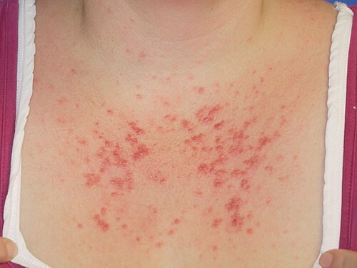
Polymorphous Light Eruption (PMLE): Key Facts at a Glance
Polymorphous light eruption (PMLE) is a common, delayed immune reaction of the skin to sunlight. It causes a very itchy rash of red bumps, blisters, or dry patches that appears hours to days after sun exposure, most often in spring and early summer. The rash usually lasts about seven to ten days and resolves without leaving permanent scars.
- Where it appears: On skin newly exposed to the sun after winter, such as the “V” of the neck, the upper chest, the forearms, and the lower legs. Rashes on the face are relatively uncommon.
- First sign: Intense itching or burning often comes before the visible skin changes.
- During a flare: Avoid further sun exposure. Otherwise the rash will worsen, spread to new areas, and last longer.
- Treatment: Topical corticosteroid creams, over-the-counter anti-itch creams, and oral antihistamines. Severe, widespread rashes may need a short course of oral corticosteroids.
- Prevention: Broad-spectrum sunscreen with SPF 50 or higher, ultraviolet-protective clothing, and avoiding the midday sun. Ultraviolet A can pass through standard window glass.
- Preventing future outbreaks: Medically supervised photohardening therapy in the early spring can desensitize the skin.
- Over the season: As the summer progresses, the skin naturally builds a tolerance that prevents further outbreaks for the rest of the season.
- Overview of Polymorphous Light Eruption (PMLE)
- Understanding the Pathophysiology of PMLE
- How Genetics Influence Polymorphous Light Eruption (PMLE)
- Frequently Asked Questions
- Scientific Sources
Overview of Polymorphous Light Eruption (PMLE)
Polymorphous Light Eruption (PMLE) is the most prevalent immunologically mediated photodermatosis worldwide, frequently described as an abnormal, delayed skin reaction to ultraviolet radiation. This condition is widely recognized by medical professionals as a seasonal, acquired, and idiopathic hypersensitivity response that most commonly occurs during the spring and early summer months when sunlight exposure significantly increases. The primary characteristic of Polymorphous Light Eruption (PMLE) is a recurrent, severely pruritic eruption that appears on the skin hours to days after sun exposure. For many patients, this condition leads to substantial discomfort, emotional distress, and a significant reduction in outdoor activities. To manage this condition effectively, one must deeply understand its complex causes, variable clinical presentations, and the broad spectrum of available treatments.
Understanding the Pathophysiology of PMLE
The exact underlying mechanisms of Polymorphous Light Eruption (PMLE) remain a subject of extensive dermatological research, though current clinical consensus identifies it as a delayed-type hypersensitivity reaction. When a susceptible individual is exposed to ultraviolet light, an endogenous photoantigen is produced or altered within the skin. In unaffected individuals, ultraviolet radiation typically induces a state of normal cutaneous immunosuppression. However, in patients suffering from Polymorphous Light Eruption (PMLE), this crucial immunosuppressive response fails. Instead of tolerating the newly formed antigens, the immune system mounts a vigorous and abnormal attack.
Recent genome-wide expression analyses have shed light on the cellular irregularities associated with this disease. Studies have highlighted that the clearance of apoptotic cells (cells programmed to die) is severely defective in patients with Polymorphous Light Eruption (PMLE). The inefficient removal of these apoptotic cells leads to the accumulation of cellular debris, which subsequently transforms into autoantigens. These autoantigens are captured by dendritic cells and presented to naive T-cells, setting off an aggressive autoimmune response. Furthermore, the over-activation of the immune system in Polymorphous Light Eruption (PMLE) involves multiple inflammatory mediators. Research indicates a significant increase in the Interleukin-1 family of cytokines, specifically IL-36 alpha and IL-36 gamma, which activate inflammatory cascades leading to the characteristic skin eruptions.
How Genetics Influence Polymorphous Light Eruption (PMLE)
Genetics undoubtedly play a significant role in the development and persistence of Polymorphous Light Eruption (PMLE). Medical literature estimates that a genetic susceptibility exists in 15 to 46 percent of cases, with many patients reporting a positive family history of the disorder. Twin studies have provided compelling evidence of a strong genetic effect, revealing a much higher concordance rate in monozygotic twins compared to dizygotic twins. Furthermore, segregation analysis suggests that up to 72 percent of the population in regions like the United Kingdom may carry a low-penetrance susceptibility allele for Polymorphous Light Eruption (PMLE). Despite the high prevalence of these genetic markers, the manifestation of the disease heavily depends on environmental triggers, specifically the intensity and duration of ultraviolet exposure.
Identifying the polymorphic light eruption Symptoms
The clinical manifestations of polymorphic light eruption are notoriously varied, which perfectly explains the term “polymorphic,” meaning “many forms”. Symptoms usually begin within a few hours to two or three days following ultraviolet exposure and can last anywhere from seven to ten days, sometimes extending up to two weeks. The most commonly reported symptom is an intense itching or burning sensation, which often precedes the visible skin changes.
“The morphology is always the same in one patient, despite the wide variety of possible presentations across different individuals.”
Common clinical morphologies of polymorphic light eruption include:
- Papular: The most frequent presentation, characterized by dense clusters of small, inflamed red or pink bumps.
- Papulovesicular: A combination of small bumps and fluid-filled blisters.
- Eczematous: Dry, red, scaling patches and plaques that closely resemble eczema.
- Erythema multiforme-like: Targetoid lesions that feature concentric rings of inflammation.
Examining polymorphous light eruption pmle Cases
In specialized clinical settings, examining diverse polymorphous light eruption pmle cases highlights the importance of patient demographics and geographic location. The prevalence of the disease is inversely related to latitude; it is remarkably higher in regions like Scandinavia, the United Kingdom, and the northern United States, while being notably lower in sun-drenched equatorial regions and Australia. In Northern Europe, the condition affects approximately 20 to 40 percent of women holidaying in the Mediterranean, whereas it only affects 1 to 5 percent of the Australasian population.
The disease predominantly affects women, with ransgender man ratio of four to one, and typical onset occurs between the ages of 20 and 40. This striking female predominance strongly suggests a hormonal component to the pathogenesis, where estrogen may act as an inhibitor of the natural ultraviolet-induced immunosuppression.
Signs of polymorphous light eruption face Rashes
While the distribution of the rash typically favors areas of the skin that are newly exposed to the sun after winter—such as the “V” of the neck, the upper chest, the extensor forearms, and the lower legs—signs of polymorphous light eruption face rashes are relatively uncommon. The face and the dorsal hands are frequently spared due to a natural phenomenon known as the skin hardening effect. Because the face is chronically exposed to sunlight throughout the year, the skin undergoes adaptive changes, including increased melanin production and a thickening of the stratum corneum. These physiological adaptations restore the normal immunosuppressive response to ultraviolet light, thereby preventing the severe inflammatory eruptions seen on normally covered body parts.
Diagnosing polymorphic light eruption Accurately
Accurately diagnosing polymorphic light eruption heavily relies on a meticulous clinical history and a thorough physical examination. A classic history of a pruritic eruption occurring hours to days after the first significant sun exposure of the spring season is a hallmark diagnostic clue. To exclude other serious photosensitive conditions, dermatologists may utilize skin biopsies, blood tests, or specialized phototesting.
| Differential Diagnosis | Distinguishing Features |
| Lupus Erythematosus | Often involves the face, persistent lesions, positive antinuclear antibodies, and positive direct immunofluorescence. |
| Solar Urticaria | Immediate onset within minutes of sun exposure and rapid resolution once exposure ceases. |
| Chronic Actinic Dermatitis | Persistent eczematous plaques with a chronic course that does not improve through seasonal photohardening. |
| Photoaggravated Atopic Dermatitis | History of chronic eczema worsened by light, rather than uniquely triggered delayed papules. |
Analyzing polymorphic light eruption pictures and Histology
When analyzing polymorphic light eruption pictures alongside histological tissue samples, medical professionals look for specific microscopic markers. The histopathology of Polymorphous Light Eruption (PMLE) is generally non-specific but remains highly supportive when correlated with clinical symptoms. Early-stage, acute lesions reveal papillary dermal edema coupled with a superficial and deep perivascular lymphocytic infiltrate.
In patients with darker skin types, a highly specific “pinpoint papular variant” is frequently observed. According to extensive studies on this variant, acute biopsies demonstrate focal vesicle formation, severe spongiosis, and red blood cell extravasation. Subacute lesions of this pinpoint variant showcase a distinct nodular collection of lymphocytes and histiocytes, accompanied by a unique claw-like extension of the epidermal rete ridges at the lateral boundaries of the lesion, which closely resembles the histology of lichen nitidus.
Understanding the polymorphous rash Variations
The variations of the polymorphous rash emphasize the critical need for personalized patient evaluation. Beyond the common papular and eczematous forms, rare triggers such as Ultraviolet C radiation have been documented in highly specific occupational settings. Welders, for instance, have been shown to develop severe polymorphous rash symptoms due to accidental Ultraviolet C overexposure when protective helmets and clothing are neglected. Phototesting in these rare cases demonstrates intense papulovesicular reactions strictly to Ultraviolet C light, proving that the photoantigen triggering the disease can be activated by different spectrums of radiation depending on the patient’s unique biological makeup.
Efficacy of Polymorphous Light Eruption (PMLE) Treatments
Effective management of Polymorphous Light Eruption (PMLE) is centered on preventing ultraviolet-induced flares, promoting natural skin tolerance, and minimizing the intense discomfort associated with active lesions. Patient education regarding sun protection is universally recognized as the foundational first-line therapy.
General preventive measures include the rigorous application of broad-spectrum sunscreens with a Sun Protection Factor of 50 or higher, specifically designed to block both Ultraviolet A and Ultraviolet B radiation. Patients must be explicitly counseled that Ultraviolet A can easily penetrate standard window glass, meaning that physical barriers, ultraviolet-protective clothing, and behavioral modifications—such as avoiding the midday sun—are absolutely mandatory.
Efficacy of polymorphous light eruption treatment Options
When preventive measures fall short, several highly effective polymorphous light eruption treatment protocols are available to manage symptoms and induce clinical remission.
For active symptom relief, dermatologists frequently prescribe potent topical corticosteroids, such as betamethasone dipropionate or mometasone ointment, for thick skin on the body, and milder hydrocortisone creams for the delicate skin of the face. Over-the-counter anti-itch creams and oral antihistamines are also highly beneficial in soothing the intense pruritus and burning sensations that accompany the eruption. For severe, widespread rashes, a short, tightly controlled course of oral corticosteroids (such as prednisolone) may be deployed to rapidly suppress systemic inflammation.
To prevent future outbreaks, controlled photohardening therapy is considered a cornerstone treatment. By exposing the patient to gradually increasing doses of Narrowband Ultraviolet B or Psoralen plus Ultraviolet A (PUVA) in the early spring, dermatologists can successfully desensitize the skin. This controlled medical exposure essentially “hardens” the skin by artificially inducing the immunosuppressive tolerance that the patient naturally lacks, providing a 65 to 100 percent photoprotection rate for the upcoming summer season.
Addressing pmle and Its Psychological Impact
Living with severe pmle often leads to profound psychosocial consequences. Patients frequently experience emotional distress, severe anxiety, depression, and social withdrawal due to the constant fear of triggering a painful flare-up. The strict requirement to avoid sunlight can drastically limit a patient’s ability to participate in outdoor recreational activities, travel, and even certain occupations. Furthermore, the extreme sun-avoidance behaviors necessitated by this condition put patients at a significant risk for Vitamin D deficiency. It is heavily recommended that individuals suffering from this disease monitor their serum Vitamin D levels and consume appropriate supplements to maintain proper bone health and systemic immunity.
In cases where phototherapy is ineffective or inaccessible, systemic pharmacological prophylaxis offers another layer of defense. Hydroxychloroquine, an antimalarial drug, is frequently prescribed as a preventive measure for patients suffering from frequent, disabling episodes. Additionally, antioxidant therapies have shown immense promise. Clinical trials have demonstrated that beta-carotene supplementation can increase a patient’s light tolerance by a factor of five, while oral administration of Polypodium leucotomos extract and nicotinamide have been proven to severely blunt the inflammatory response prior to sun exposure.
Frequently Asked Questions
What is polymorphous light eruption?
Polymorphous light eruption is a highly common, delayed immune reaction of the skin triggered by exposure to sunlight or artificial ultraviolet radiation. It manifests as a severely itchy and uncomfortable rash featuring red bumps, blisters, or dry patches that typically appear hours to days after sun exposure. The condition predominantly affects individuals during the spring and early summer when the skin is unaccustomed to intense ultraviolet rays, and it arises due to an abnormal immune system response that fails to tolerate natural light-induced skin changes.
How to treat polymorphic light eruption?
The most effective way to treat an active flare involves minimizing further sun exposure and applying topical corticosteroid creams to rapidly reduce inflammation and itching. For severe episodes, doctors may prescribe a short course of oral steroids or recommend oral antihistamines to alleviate intense discomfort. To prevent future outbreaks, patients are advised to strictly use broad-spectrum sunscreens, wear protective clothing, and undergo medically supervised photohardening therapy (such as Narrowband UVB) in the early spring to desensitize the skin to ultraviolet radiation.
How long does polymorphic light eruption last?
Once the skin is exposed to the triggering ultraviolet light, the resulting rash usually persists for about seven to ten days, provided that the affected individual strictly avoids any further sun exposure. If the patient continues to expose the inflamed skin to sunlight, the eruption will actively worsen, spread to new areas, and last significantly longer. Fortunately, the rash ultimately resolves on its own without leaving any permanent scars, and as the summer progresses, the skin naturally builds a tolerance that prevents further outbreaks for the remainder of the season.
The following posts may interest you
Sun Poisoning Guide: Symptoms, Treatment & Prevention
Best Sunscreen for Babies: Safe & Gentle UV Protection
How to Protect Baby from Sun in Summer: Safe & Simple Tips
Disclaimer: The content provided on BabyKidsHealth.com is for informational purposes only and is not intended as a substitute for professional medical advice, diagnosis, or treatment. Always seek the advice of your physician or other qualified health provider with any questions you may have regarding a medical condition.
Sources
Polymorphic light eruption (NHS, UK)
https://www.nhs.uk/conditions/polymorphic-light-eruption/
Polymorphous light eruption (MedlinePlus, U.S. National Library of Medicine)
https://medlineplus.gov/ency/article/001477.htm
Polymorphous Light Eruption– An Indian Scenario
https://pmc.ncbi.nlm.nih.gov/articles/PMC8088173
Polymorphous light eruption
https://onlinelibrary.wiley.com/doi/abs/10.1111/j.1600-0781.2008.00343.x
Polymorphous light eruption: A clinical, photobiologic, and follow-up study of 110 patients
https://www.sciencedirect.com/science/article/abs/pii/S0190962200701022
Polymorphous light eruption: Action spectrum and photoprotection
https://www.sciencedirect.com/science/article/abs/pii/S0190962286700881
Psychologic distress in polymorphous light eruption and its relationship to patients’ beliefs about their condition
https://www.sciencedirect.com/science/article/abs/pii/S0190962206028684
Polymorphous light eruption: Experimental reproduction of skin lesions
https://www.sciencedirect.com/science/article/abs/pii/S0190962282800182
Polymorphous Light Eruption: a Review
https://link.springer.com/article/10.1007/s13671-019-0264-y
Skin microbiome and polymorphous light eruption
https://academic.oup.com/bjd/article-abstract/192/4/569/7933263