- Comprehensive Guide to Managing Impetigo
- Pathophysiology and Epidemiology of Impetigo
- Effective Strategies
- Frequently Asked Questions
- Scientific Sources
Comprehensive Guide to Managing Impetigo
Pediatric health requires constant vigilance, especially concerning highly contagious dermatological conditions. Impetigo stands out as one of the most prevalent bacterial skin infections affecting young children, typically between the ages of two and five. This condition manifests as a superficial infection that can rapidly spread among siblings, classmates, and caregivers if proper interventions are not established promptly. It is crucial to ground our understanding of this condition in scientific literature, ensuring that evidence-based practices are implemented to promote swift recovery and prevent recurrence.
As we delve into the pathophysiology, clinical presentations, and management strategies for this condition, it is vital to remember that child skin is uniquely susceptible to disruptions in its barrier function. Micro-traumas from insect bites, eczema excoriations, or minor abrasions often serve as the entry point for the causative pathogens. The primary bacteria responsible for this infection are Staphylococcus aureus and Streptococcus pyogenes. Timely recognition and appropriate management are absolutely essential to prevent complications and limit the spread of the infection within the community.
Pathophysiology and Epidemiology of Impetigo
The human skin is host to a variety of resident microflora that generally protect against pathogenic invasion. However, transient flora, including pathogenic strains of bacteria, can colonize the skin when the natural barrier is compromised. The process of colonization involves the irreversible adherence of bacteria to specific receptors on the host cells. Research indicates that the availability of fibronectin receptors, often exposed due to minor skin trauma, facilitates the adherence of pathogenic bacteria.
The primary bacterial agents responsible for this infection are Staphylococcus aureus and group A beta-hemolytic Streptococcus (GABHS), or a combination of both. In recent decades, Staphylococcus aureus has emerged as the dominant pathogen in most developed regions. Transmission occurs primarily through direct skin-to-skin contact. The highly contagious nature of the bacteria means that autoinoculation is common; children often spread the bacteria from one part of their body to another by scratching. Furthermore, sharing towels, bedding, clothing, or toys can facilitate the spread among family members and within childcare facilities.
Diagnosing Impetigo Correctly
Diagnosis is predominantly clinical, based on the characteristic appearance of the honey-colored crusts or flaccid blisters. Routine bacteriological swabs are generally unnecessary for standard cases. However, in instances where the infection fails to respond to initial empirical therapy, or during an outbreak, swabbing the exudate beneath the crust can be valuable for identifying the specific pathogen and determining its antibiotic susceptibility.
Recognizing impetigo symptoms early
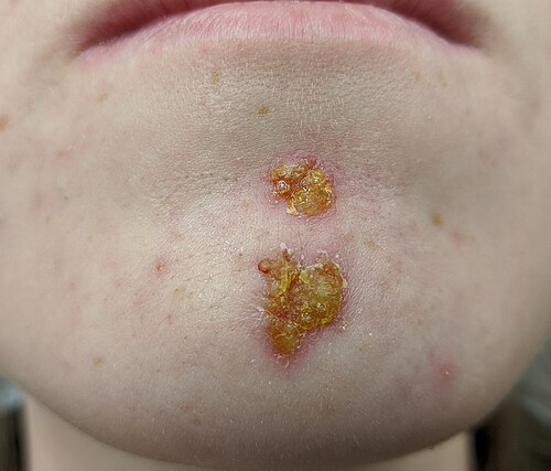
Non-bullous Impetigo accounts for approximately 70% of all cases. It typically begins on skin that has been traumatized, such as by insect bites, abrasions, or underlying conditions like varicella or eczema. The infection initially presents as tiny vesicles or pustules that rapidly rupture. Once ruptured, they leave behind superficial erosions covered by a characteristic honey-colored crust. These impetigo symptoms are generally less than 2 centimeters in diameter and are associated with minimal pain, though intense itching is common. Regional lymphadenopathy (swollen lymph nodes) is frequently observed in patients. Without treatment, these lesions may slowly spread over several weeks, but they usually resolve without leaving permanent scars.
Managing bullous impetigo variants
Bullous impetigo is exclusively caused by Staphylococcus aureus and represents a localized form of staphylococcal scalded skin syndrome. Unlike the non-bullous variant, it often develops on intact skin. This variant is frequently found on the trunk, axillae, extremities, and in the diaper area of infants.
Analyzing impetigo images for clues
Clinical impetigo images of the bullous variant typically reveal large, fragile, flaccid bullae (blisters) that contain clear or yellow fluid. These blisters are prone to rupturing easily. Following rupture, a characteristic collarette of scale forms at the periphery, leaving a shallow, moist erosion with a thin brown crust. Caregivers and healthcare professionals often rely on the distinct visual presentation seen in these cases to differentiate the condition from other blistering disorders like thermal burns or contact dermatitis.
When an impetigo rash begins to spread, it is usually a sign of unchecked bacterial proliferation and autoinoculation. Parents and caregivers must act swiftly to cover the affected areas loosely and enforce strict hand hygiene to halt the spread of the impetigo rash.
Effective Strategies
The primary goals of therapy are to relieve discomfort, improve cosmetic appearance, prevent the spread of the organism, and avoid rare complications. Treatment selection depends on the extent of the infection and local antibiotic resistance patterns.
Proper wound care is the cornerstone of healing. Gently washing the sores with mild soap and warm water to remove crusts allows topical treatments to penetrate effectively and accelerates the healing process.
Optimizing Impetigo Treatment Protocols
For localized, uncomplicated infections, topical therapies are highly effective and are generally preferred over systemic treatments. Topical applications minimize systemic absorption, thereby reducing the risk of adverse gastrointestinal side effects and lowering the potential for systemic antibiotic resistance. A successful impetigo treatment plan often begins with meticulous hygiene and the application of targeted antibacterial ointments.
Formulating a plan for treatment impetigo
When deciding on the best course of action for treatment impetigo, healthcare providers must consider the patient’s age, the extent of the lesions, and any underlying health conditions. Children with active infections should be excluded from school or childcare until they have completed at least 24 to 48 hours of appropriate antibiotic therapy, or until the sores have entirely crusted over and healed.
Exploring impetigo antibiotics options
Several topical impetigo antibiotics have demonstrated high efficacy. Mupirocin ointment is a widely prescribed option that effectively targets both Staphylococcus aureus and Streptococcus pyogenes. It is typically applied three times daily for a period of five to ten days. Fusidic acid is another excellent topical agent utilized extensively in many countries, showing comparable efficacy to mupirocin. Additionally, retapamulin, a newer pleuromutilin antibiotic, has been approved for short-course topical therapy and provides a valuable alternative, especially given the rising concerns about bacterial resistance.
Considering systemic Impetigo therapies
Oral antibiotics are indicated for patients with widespread lesions, bullous variants, or when topical therapy is impractical. Beta-lactam antibiotics, such as cephalexin or amoxicillin/clavulanate, remain appropriate first-line empirical choices for systemic therapy. In regions with a high prevalence of community-associated methicillin-resistant Staphylococcus aureus (CA-MRSA), alternative oral agents like clindamycin, trimethoprim-sulfamethoxazole, or doxycycline may be necessary based on local susceptibility data. Penicillin and standard erythromycin are no longer recommended due to widespread resistance.
| Preventive Action | Description | Frequency |
| Handwashing | Use soap and warm water thoroughly, especially after touching wounds. | Frequently |
| Wound Care | Clean minor cuts, scrapes, and insect bites immediately. | As needed |
| Nail Trimming | Keep child’s fingernails short to prevent skin damage from scratching. | Weekly |
| Linen Hygiene | Wash towels, bed sheets, and clothes in hot water. Do not share. | Daily during infection |
Maintaining the integrity of a child’s skin goes beyond treating the immediate infection. Preventing future episodes of Impetigo requires a holistic approach to skin care. Children with underlying dermatological conditions, such as atopic dermatitis or eczema, are particularly vulnerable because their skin barrier is chronically compromised. In these cases, regular use of emollients and gentle, fragrance-free cleansers can fortify the skin’s natural defenses, making it more difficult for bacteria to take hold. Furthermore, educating children about the importance of not sharing personal items at school or daycare is a crucial step in minimizing outbreaks.
As caregivers, remaining vigilant and proactive when the first signs of Impetigo appear will ensure that our communities remain healthier and our children can play, learn, and grow without the discomfort of recurrent skin infections. Careful monitoring and adherence to prescribed medical regimens are the most reliable pathways to fully eradicating the bacteria and restoring the skin’s natural, healthy state. Impetigo is a very manageable condition when approached with the right knowledge and tools, ensuring that pediatric patients recover swiftly and safely.
Frequently Asked Questions
What is impetigo?
It is a highly contagious bacterial skin infection most commonly affecting infants and young children. It manifests primarily as red sores that quickly rupture, ooze for a few days, and then form a characteristic yellowish-brown or honey-colored crust. The infection targets the superficial layers of the epidermis and is generally caused by staphylococcal or streptococcal bacteria.
How to get rid of impetigo in 24 hours?
It is medically impossible to completely cure the infection within 24 hours, as the body and antibiotics require time to eradicate the bacteria and heal the skin. However, initiating treatment with a prescribed topical or oral antibiotic can stop the progression and reduce contagiousness within the first 24 to 48 hours. Consistent application of prescribed medication and meticulous hygiene are the fastest paths to full recovery.
Is impetigo contagious?
Yes, it is extremely contagious and spreads very easily through direct skin-to-skin contact with infected individuals. It can also be contracted indirectly by touching items that an infected person has recently used, such as towels, clothing, bedding, or toys. The condition remains contagious until the sores are completely healed or the patient has been undergoing appropriate antibiotic therapy for at least 24 to 48 hours.
What does impetigo look like?
The visual presentation varies depending on the specific type of infection. The most common non-bullous form looks like clusters of red, itchy sores that burst and leave behind a thick, golden or honey-colored crust. The less common bullous form appears as large, fluid-filled blisters that are fragile, often breaking to leave raw, red skin and a thin brown crust at the edges.
How do you get impetigo?
You contract the infection when the causative bacteria, typically Staphylococcus aureus or Streptococcus pyogenes, enter the body through a break in the skin. This break can be as minor as a scratch, an insect bite, or irritated skin from conditions like eczema or a runny nose. Direct contact with someone who has the infection or sharing their personal items greatly increases the likelihood of the bacteria colonizing these minor skin abrasions.
The following posts may interest you
Poison Ivy Rash: Causes, Symptoms, and Best Treatments
Cradle Cap Guide: Causes, Symptoms, and Treatments
Molluscum Contagiosum: Symptoms, Causes & Treatments
Sources
Impetigo: An Overview
https://onlinelibrary.wiley.com/doi/abs/10.1111/j.1525-1470.1994.tb00092.x
Impetigo: Diagnosis and Treatment
https://www.aafp.org/afp/2014/0815/p229
A systematic review and meta-analysis of treatments for impetigo
https://pmc.ncbi.nlm.nih.gov/articles/PMC1314624
Impetigo – review
https://www.scielo.br/j/abd/a/cpm9SxPg4rwGjq7Qf679Ffq/?lang=en
Diagnosis and Treatment of Impetigo
https://www.aafp.org/afp/2007/0315/p859
Double-blind study comparing erythromycin and mupirocin for treatment of impetigo in children: implications of a high prevalence of erythromycin-resistant Staphylococcus aureus strains
https://journals.asm.org/doi/abs/10.1128/aac.36.2.287
The Importance of Scabies Coinfection in the Treatment Considerations for Impetigo
Natural history of impetigo: II. Etiologic agents and bacterial interactions
https://www.jci.org/articles/view/107109
Streptococcal impetigo and acute glomerulonephritis in children in Cairo
https://academic.oup.com/bjd/article-abstract/98/1/53/6670007
image:
By James Heilman, MD – Own work, CC BY-SA 4.0, https://commons.wikimedia.org/w/index.php?curid=90030642