- Virology and Molecular Biology of Rhinovirus
- Pathogenesis and Epithelial Barrier Dynamics of Rhinovirus
- Clinical Manifestations and High-Risk Populations
- Rhinovirus and Pediatric Asthma Pathogenesis
- Transmission Dynamics and Infection Control
- Diagnostic Approaches for Rhinovirus
- Current Management and Investigational Therapies
- Clinical Red Flags and Caregiver Guidance
- Frequently Asked Questions
- Scientific Sources
Rhinovirus: Comprehensive Clinical Guide to Pediatric and Adult Respiratory Illness
Understanding the pervasive biological behavior of Rhinovirus is essential for clinical practitioners, pediatric specialists, and caregivers who manage acute upper and lower respiratory conditions. As a primary viral pathogen responsible for the vast majority of mild colds, Rhinovirus exhibits complex transmission pathways, diverse serotypic variants, and substantial immune-modulating properties within respiratory tissues. While a standard viral episode caused by Rhinovirus frequently resolves spontaneously without specialized therapeutic intervention, vulnerable populations—such as young infants, preterm newborns, and children diagnosed with reactive airway diseases—face substantial risks of prolonged airway impairment, persistent wheezing episodes, and sudden physiological decompensation.
1. Virology and Molecular Biology of Rhinovirus
Viral Structure and Genomic Organization
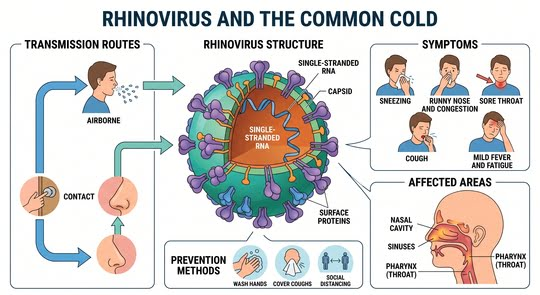
Human rhinoviruses are non-enveloped, positive-sense, single-stranded RNA pathogens belonging to the family Picornaviridae. The outer protein shell forms an icosahedral architecture constructed from sixty repeating copies of four distinct structural viral capsid proteins: VP1, VP2, VP3, and VP4. Structural proteins VP1 through VP3 form the exposed external surface of the virion, establishing prominent topography characterized by deep surface depressions known as canyons. The smaller, largely internal protein VP4 anchors the viral RNA complex within the core and plays a decisive role during host cell attachment, receptor binding, and subsequent genomic release.
Because Rhinovirus possesses an RNA-dependent RNA polymerase that completely lacks proofreading activity, genomic replication produces continuous antigenic drift. This mechanism underlies the staggering antigenic diversity found across more than 160 recognized serotypes. Genetic cataloging classifies the pathogen into three distinct species:
- HRV-A: The most historically recognized group alongside HRV-B, associated with traditional seasonal rhinorrhea, pharyngeal irritation, and moderate respiratory illness.
- HRV-B: Genetically distinct from HRV-A, generally associated with milder symptomatic courses, though fully capable of replicating within bronchial epithelium.
- HRV-C: Identified through contemporary molecular sequencing techniques, HRV-C possesses distinct biological receptors and demonstrates an affinity for lower bronchial structures, precipitating severe wheezing attacks and sudden asthma exacerbations in pediatric hosts.
The cellular receptors mediating viral internalization vary across strains. Over 90% of HRV-A and HRV-B isolates—collectively designated the major receptor group—bind directly to Intercellular Adhesion Molecule-1 (ICAM-1) located on respiratory epithelial cells. A smaller subset of serotypes designated the minor receptor group gains cellular entry via members of the low-density lipoprotein (LDL) receptor family. In contrast, HRV-C relies upon Cadherin-related family member 3 (CDHR3) for host attachment. Following binding, conformational alterations in capsid structure cause shedding of VP4 and externalization of the hydrophobic amino-terminal domains of VP1, permitting the single-stranded RNA genome to cross host cell membranes into the cytoplasm to initiate rapid viral polyprotein translation.
The Genetic Continuum of Rhinovirus Enterovirus Lineages
The clinical diagnosis and molecular characterization of respiratory illness regularly intersect at the genomic boundary between Rhinovirus and non-polio enteroviruses. Belonging to the shared Picornaviridae family, rhinovirus enterovirus classifications share structural symmetries, conserved genomic regions, and overlapping seasonal prevalence patterns. Standard multiplex real-time polymerase chain reaction (RT-PCR) panels target highly conserved 5′ untranslated regions, which frequently report a combined Rhinovirus enterovirus positive result because differentiating both agents requires labor-intensive nested PCR and genetic sequencing.
Despite structural and genomic similarities, critical clinical distinctions exist between these viral groups:
- Human rhinovirus replicates optimally at temperatures between 33°C and 35°C—the typical physiological microenvironment of nasal and upper pharyngeal passages—though sustained core temperatures of 37°C in the tracheobronchial tree do not prevent viable lower airway replication.
- Enteroviruses demonstrate absolute acid stability, allowing safe gastrointestinal transit, fecal shedding, and systemic hematogenous dissemination to secondary organ systems including the liver, cardiac muscle, and the central nervous system.
- Rhinovirus exhibits marked acid lability, rendering the virion inactive when exposed to low pH conditions below 5.0 to 6.0. However, viable viral particles and viral genetic transcripts are regularly detected in rectal swabs of young children, illustrating that swallowed respiratory secretions survive intestinal transit under certain physiological states.
2. Pathogenesis and Epithelial Barrier Dynamics of Rhinovirus
Disruption of Airway Integrity Without Overt Cytopathology
Unlike aggressive respiratory viruses such as influenza or respiratory syncytial virus (RSV) that trigger extensive cytolytic necrosis, cellular sloughing, and denudation of the respiratory mucosa, Rhinovirus maintains an intriguing host relationship characterized by minimal microscopic cytopathology. Biopsies of nasal and bronchial tissues infected with Rhinovirus exhibit structurally intact cellular monolayers. The clinical manifestation of infection arises from host-directed immune signaling and selective disassembly of apical barrier junctions rather than physical viral destruction of the airway mucosa.
Upon productive infection of upper or lower airway epithelium, Rhinovirus triggers a rapid downregulation and mechanical fragmentation of vital tight junction complexes. Specifically, transcription and membrane stability of:
- Zona occludens-1 (ZO-1),
- Claudin-1, and
- E-cadherin
decline precipitously within 24 to 48 hours. This localized junctional dissolution increases vascular and mucosal permeability, allowing massive exudation of protein-rich plasma into the airway lumen. Simultaneously, broken intercellular barriers expose basolateral pattern recognition receptors (PRRs) and provide environmental aeroallergens direct access to submucosal immune sentinels.
| Sequential Phase | Biological Action | Microscopic and Physiological Consequence |
| Phase 1: Inhalation & Binding | Viral virion attaches to surface receptors (ICAM-1, LDL, or CDHR3). | Localized engagement along mucosal surface without direct tissue damage. |
| Phase 2: Cellular Entry & Replication | Viral genomic RNA enters host cytoplasm and initiates non-cytolytic replication. | Intact cell morphology maintained without cytolytic sloughing or cell death. |
| Phase 3: Junctional Breakdown | Downregulation of apical tight junctions (ZO-1, Claudin-1, E-cadherin). | Intercellular cleft opening and compromise of airway epithelial barrier integrity. |
| Phase 4: Dual Clinical Cascade | Epithelial permeability increases alongside basolateral receptor exposure. | Profuse rhinorrhea, mucosal edema, and access for environmental allergens. |
Innate Cytokine Release and Granulocyte Recruitment
The human immune response to Rhinovirus initiates almost instantly upon viral entry. Host cells identify viral architecture through several coordinated PRRs:
- Surface-bound Toll-Like Receptor 2 (TLR2) detects external viral capsid sequences.
- Endosomal Toll-Like Receptors 3, 7, and 8 (TLR3, TLR7, TLR8) detect double-stranded replicative intermediates and single-stranded viral RNA.
- Cytoplasmic helicases, including Retinoic Acid-Inducible Gene I (RIG-I) and Melanoma Differentiation-Associated Protein 5 (MDA-5), bind intracellular replicating viral RNA.
Activation of these intracellular signaling cascades prompts the rapid synthesis and mucosal secretion of type I (interferon-alpha, interferon-beta) and type III interferons (interleukin-28, interleukin-29). Paradoxically, along with protective interferons, airway epithelium releases an array of potent pro-inflammatory chemokines and cytokines, including CXCL8 (interleukin-8), CXCL5, CXCL10, interleukin-6, and interleukin-11. Interleukin-8 initiates a massive influx of circulating neutrophils into the submucosa and nasal passages. Neutrophils discharge active myeloperoxidase and tissue elastases that drive thick purulent exudate formation. Furthermore, viral activation stimulates tissue kallikreins, converting systemic kininogens into bradykinin and lysyl-bradykinin. The accumulation of these kinins directly stimulates sensory C-fibers, triggering severe sore throat pain, hyperesthetic pharyngeal tickling, and reflex sneezing episodes.
3. Clinical Manifestations and High-Risk Populations
Clinical Spectrum Across Diverse Age Groups
The symptomatic expression of Rhinovirus varies significantly according to patient age, baseline airway caliber, and the underlying competency of mucosal immunity:
| Clinical Parameter | Neonates and Young Infants | Toddlers and Older Children | Healthy Adults | High-Risk and Immunocompromised Hosts |
| Typical Incubation | 1 to 3 days | 1 to 3 days | 1 to 3 days | 1 to 2 days |
| Dominant Early Signs | Poor feeding, nasal snorting, lethargy, low fever | Copious rhinorrhea, sneezing, paroxysmal cough | Pharyngeal soreness, sneezing, nasal stuffiness | Rapid lower airway cough, wheezing, dyspnea |
| Secretions Profile | Clear changing rapidly to tenacious obstruction | Watery to thick mucopurulent discharge | Watery rhinorrhea transitioning to thick mucus | Profuse secretions, alveolar exudative debris |
| Lower Airway Risks | Apnea, bronchiolitis, sudden collapse | Wheezy bronchitis, reactive airway spasm | Transient bronchial hyperreactivity | Viral pneumonitis, diffuse alveolar damage |
| Resolution Timeline | 10 to 14 days | 7 to 10 days | 5 to 7 days | 2 to 6 weeks (protracted viral shedding) |
In daily pediatric practice, evaluating rhinovirus symptoms requires careful distinction between uncomplicated upper tract coryza and progressive lower tract compromise. While adults experience headaches, modest malaise, and rhinorrhea without significant pulmonary decrement, the clinical presentation shifts dramatically across pediatric demographics.
An active rhinovirus baby infection frequently manifests with profound nutritional difficulties. Because young infants are obligate nasal breathers, even moderate nasal mucosal congestion interferes with nursing, causing fatigue, poor fluid intake, and rapid dehydration. In rhinovirus in infants, localized swelling can rapidly induce significant tachypnea and subtle inspiratory stridor.
When analyzing rhinovirus in children, clinicians observe that younger demographics display higher fevers than adults, frequently exceeding 38.5°C during the initial 48 hours. Furthermore, rhinovirus in childrens cohorts attending group daycare centers often presents with concurrent bilateral middle ear pressure, acute otitis media, and periorbital edema. Parents of a child with rhinovirus in kids settings report persistent daytime coughing that intensifies when supine due to posterior pharyngeal drainage.
Among mobile age brackets, identifying rhinovirus in toddlers involves tracking irritability, refusal of solids, hoarseness, and sudden wheezing episodes. Because maternal antibody concentrations wane significantly after six months of age, rhinovirus in babies between 6 and 24 months represents the highest demographic for pediatric emergency evaluations and hospitalizations.
Clinical Warning: Sudden onset of tachypnea, intercostal retractions, or feeding exhaustion in young infants warrants immediate medical evaluation to rule out acute viral bronchiolitis or reactive airway failure.
Severe Complications in At-Risk Pediatric Cohorts
While most infections follow a self-limited course, severe complications can emerge in specific vulnerable pediatric populations:
Premature Infants and Bronchopulmonary Dysplasia
Infants born prematurely lack late-gestation transplacental transfers of maternal immunoglobulin G. When infected with Rhinovirus, their structurally smaller airways and immature lung compliance make them susceptible to acute bronchiolitis, respiratory syncytial virus co-infections, and sudden hypoxic episodes requiring invasive mechanical ventilation.
Children with Underlying Asthma and Atopic Diathesis
Rhinovirus is the primary driver of viral-induced pediatric asthma attacks, accounting for 60% to 80% of all childhood asthma hospitalizations. When Rhinovirus disrupts the bronchial epithelium of an atopic child, elevated local levels of Interleukin-8 and eosinophilic cationic proteins synergize with pre-existing immunoglobulin E (IgE) responses. This dual activation provokes severe bronchospasm, mucous plugging, and extended oxygen dependence.
Immunocompromised Patients
Children receiving cytotoxic chemotherapy, congenital immunodeficiency patients, and pediatric stem-cell transplant recipients experience high rates of lower respiratory parenchymal invasion. Rather than clearing the viral load within 10 days, these patients may exhibit continuous lower airway viral replication for weeks, predisposing them to viral pneumonitis, diffuse alveolar damage, and secondary bacterial infections caused by Streptococcus pneumoniae or Staphylococcus aureus.
Central Nervous System Complications and SIDS
Although historically considered non-invasive, Rhinovirus has been documented invading non-respiratory sites. Clinical literature has linked Rhinovirus infections to rare cases of pediatric encephalitis, sudden neurological deterioration, and fatal outcomes classified under Sudden Infant Death Syndrome (SIDS).
In a notable medical case, a 20-day-old infant who presented with mild rhinitis died unexpectedly during sleep. Comprehensive postmortem molecular evaluations ruled out trauma and common bacterial pathogens. RT-PCR testing confirmed the presence of Rhinovirus type A10 within the lungs, rectal mucosa, and cerebrospinal fluid (CSF). Autopsy findings showed:
- Histiocytic alveolitis,
- Interstitial pulmonary edema,
- Direct viral antigens inside alveolar macrophages, and
- Abnormally elevated protein and lactate levels within the cerebrospinal fluid.
This case demonstrates that Rhinovirus can cross the blood-brain barrier—potentially via hematogenous routes or direct retrograde migration along the olfactory nerve tract—triggering inflammatory cytokine storms that impair central autonomic cardiorespiratory regulation in young infants.
4. Rhinovirus and Pediatric Asthma Pathogenesis
The Causation Versus Unmasking Dilemma
The exact relationship between Rhinovirus infections in early life and the subsequent development of chronic childhood asthma remains an area of ongoing investigation. Two primary hypotheses explain the strong epidemiological correlation observed between early viral wheezing and long-term airway hyperresponsiveness:
- The Causative Hypothesis (Structural Damage): Repeated severe lower respiratory infections caused by Rhinovirus disrupt delicate basement membranes during periods of rapid alveolar and bronchial morphogenesis. Resulting fibroblastic proliferation, subepithelial thickening, and chronic airway remodeling permanently alter airway compliance, predisposing the child to persistent asthma.
- The Epiphenomenon Hypothesis (Pre-existing Vulnerability): Infants who develop wheezing when exposed to Rhinovirus possess innate anatomical and immunological vulnerabilities. Congenitally smaller airway calibers, deficient native interferon production (such as impaired interferon-beta or interleukin-15 expression), and genetic polymorphisms within the 17q21 locus or protocadherin-1 genes predispose these children to both severe symptomatic viral infections and chronic asthma.
| Model Component | Causative Remodeling Mechanism | Unmasked Susceptibility Mechanism |
| Primary Driver | Recurrent lower tract epithelial damage induced by severe viral episodes. | Inherent host vulnerabilities, including small baseline airways and 17q21 gene variants. |
| Airway Impact | Epithelial disruption, subepithelial fibrosis, and impaired alveolar development. | Early exposure of basolateral receptors and deficient innate interferon release. |
| Synergy Factor | Viral replication accelerates physical basement membrane remodeling. | Concomitant allergen sensitization triggers hyperreactive IgE-mediated cascades. |
| Clinical Outcome | Structural airway narrowing and progressive fixed bronchial hyperresponsiveness. | Frequent episodic wheezing with high probability of early school-age asthma. |
Longitudinal birth cohort data, including findings from the Childhood Origins of Asthma (COAST) study, demonstrate that infants who experience moderate-to-severe Rhinovirus wheezing episodes during the first three years of life have an odds ratio greater than 25 for developing doctor-diagnosed asthma by six years of age. When infant aeroallergen sensitization occurs alongside recurrent viral infections, the risk of developing chronic atopic asthma multiplies substantially.
5. Transmission Dynamics and Infection Control
Viral Shedding and Environmental Stability
Controlling the spread of Rhinovirus requires understanding its environmental resilience. As a non-enveloped viral agent, Rhinovirus displays high structural resistance to environmental desiccation and temperature fluctuations compared to enveloped viruses like influenza or RSV.
The primary modes of viral spread include:
- Direct Hand Contact: Transfer of viral-laden nasal secretions through skin-to-skin touch, followed by autoinoculation into nasal passages or ocular conjunctiva.
- Contaminated Fomites: The pathogen remains viable on smooth household surfaces (such as plastic toys, doorknobs, and countertops) for up to several hours to several days, facilitating high secondary attack rates in households and daycares.
- Aerosol Transmission: Airborne droplets expelled during paroxysmal coughing or sneezing remain suspended and can initiate infection upon direct inhalation into the upper or lower respiratory tree.
Understanding the rhinovirus infectious period is necessary for implementing effective infection control measures in home and school settings. Symptomatic individuals shed the highest quantities of viable virus during the first 48 to 72 hours following symptom onset, aligning with peak nasal discharge and sneezing frequency. However, quantitative PCR analyses confirm that viral RNA shedding regularly continues for 10 to 14 days in healthy individuals, and can persist for up to 30 days in young children and infants. In immunocompromised patients, low-grade shedding may continue for several months. Consequently, individuals can spread Rhinovirus even as their clinical symptoms improve.
6. Diagnostic Approaches for Rhinovirus
Laboratory Identification and Clinical Differentiation
In primary care settings, uncomplicated upper respiratory episodes rarely require definitive diagnostic testing. Experienced clinicians diagnose viral colds based on clinical presentation, including clear nasal secretions, mild pharyngeal erythema, low-grade temperature elevations, and absence of lower lung crackles. However, in emergency departments, intensive care environments, and specialized pediatric clinics, determining the etiology of severe respiratory illness requires specific diagnostic methods:
| Diagnostic Technique | Practical Application | Analytical Strengths | Clinical Limitations |
| Multiplex Real-Time RT-PCR | Standard testing for hospitalized children and high-risk respiratory cases. | Rapid turnaround (hours), high analytical sensitivity, pan-serotype detection. | May detect asymptomatic carriage, resolving RNA fragments, or report combined RV/EV results. |
| Direct Immunofluorescence (DFA) | Rapid clinical antigen screening in emergency settings. | Good specificity for active viral replication in nasal washings. | Significantly lower analytical sensitivity compared to modern PCR methods. |
| Viral Cell Culture | Specialized epidemiological research and surveillance studies. | Conclusive proof of actively replicating, viable infectious virions. | Labor-intensive, requires 3 to 7 days, and fails to support difficult fastidious serotypes. |
- Multiplex Real-Time RT-PCR: The clinical gold standard for detecting respiratory pathogens. These diagnostic panels provide high analytical sensitivity and deliver rapid results within hours. Because genetic targets cross-amplify across the Enterovirus genus, positive results are frequently reported as Rhinovirus enterovirus.
- Direct Immunofluorescence Assays (DFA): DFA uses fluorescently labeled antibodies to detect viral capsid antigens in nasopharyngeal aspirates. While offering higher clinical specificity for active high-titer viral replication, DFA exhibits lower sensitivity than nucleic acid amplification methods.
- Viral Cell Culture: Historically considered the reference method, isolating Rhinovirus in cell cultures (such as human embryonic lung fibroblasts or HeLa cells) requires specialized laboratory expertise, incubation at lower temperatures (33°C), and 3 to 7 days of processing time. Culture is impractical for timely clinical decision-making and is primarily reserved for research applications.
When analyzing respiratory specimens, medical providers must recognize that a positive RT-PCR result does not definitively establish that Rhinovirus is the primary driver of an acute illness. Because low levels of viral RNA can linger in the nasopharynx for weeks following a cold, sensitive molecular panels can detect asymptomatic colonization or remnants of previous infections. Clinicians must correlate viral detection with physical findings, oxygen saturation metrics, and markers of systemic inflammation.
7. Current Management and Investigational Therapies
Evidence-Based Supportive Care
At present, there is no approved antiviral medicine or vaccine designed to cure or prevent illness caused by Rhinovirus. Consequently, clinical management relies entirely upon targeted supportive care, prevention of dehydration, and close respiratory monitoring:
- Airway Hydration and Saline Irrigation: Instilling isotonic or hypertonic saline drops into the nasal cavity softens crusts and clears thick mucus. In young infants, gentle bulb suctioning or mechanical aspirators before feedings relieves obstruction, restores obligate nasal breathing, and prevents nutritional compromise.
- Environmental Air Humidification: Using cool-mist room humidifiers maintains mucosal hydration, prevents epithelial drying from indoor air conditioning or winter heating, and eases nocturnal coughing paroxysms. Warm-mist vaporizers should be avoided in pediatric spaces due to scald risks.
- Adequate Fluid Intake: Clinicians should instruct caregivers to provide frequent, small-volume feedings of breast milk, infant formula, or water. Adequate systemic hydration thins secretions, supports mucous clearance, and maintains normal renal perfusion.
- Targeted Antipyretics and Analgesics: When systemic fever or headache causes marked distress, acetaminophen or ibuprofen may be administered following weight-based dosing guidelines. Ibuprofen should only be given to infants older than six months of age who maintain adequate oral hydration. Acetylsalicylic acid (aspirin) must be avoided in infants, children, and teenagers with viral infections to eliminate the risk of Reye syndrome.
- Honey for Cough Relief in Children Over 12 Months: For pediatric patients older than one year, administering 2.5 to 5 milliliters of pure buckwheat or wildflower honey provides soothing demulcent activity, reducing nocturnal cough severity. Honey is contraindicated in infants under 12 months due to the risk of infant botulism caused by Clostridium botulinum spores.
Caregiver Management Summary: Optimal supportive management centers on proactive airway clearance using saline drops, continuous cool-mist humidification, and close monitoring of hydration via daily diaper output.
Safety Warning: Over-the-counter multi-symptom cold and cough preparations containing decongestants, antihistamines, or antitussives should never be administered to children under two years of age. These compounds offer no established clinical benefit and carry serious risks of severe adverse reactions, including cardiac arrhythmias, depressed consciousness, and fatal toxicity.
Pediatric Antipyretic and Supportive Regimen Reference
The following table summarizes established clinical guidelines for fever and symptom management in pediatric populations:
| Medication / Intervention | Approved Age Range | Standard Pediatric Dosing | Key Clinical Considerations |
| Acetaminophen | Neonates to Adolescents | 10–15 mg/kg per dose every 4–6 hours (Max: 75 mg/kg/day) | Preferred antipyretic for infants under 6 months; avoids renal stress. |
| Ibuprofen | Infants $\ge$ 6 Months | 5–10 mg/kg per dose every 6–8 hours (Max: 40 mg/kg/day) | Requires adequate oral intake; contraindicated in dehydration or renal compromise. |
| Nasal Saline & Suction | All Ages (Birth+) | 2–3 drops per nostril before feedings and sleep | Mechanical suctioning must be gentle to prevent mucosal edema and trauma. |
| Cool-Mist Humidifier | All Ages (Birth+) | Continuous in sleep environment | Requires daily cleaning and water replacement to avoid microbial growth. |
| Natural Honey | Children $\ge$ 12 Months | 2.5–5 mL (half to one teaspoon) as needed before sleep | Strictly contraindicated under 1 year due to infant botulism risks. |
Inappropriate Therapies and Antiviral Pipelines
A critical responsibility in outpatient medicine is preventing inappropriate antimicrobial use. Because Rhinovirus is an RNA virus lacking peptidoglycan walls or bacterial metabolic pathways, antibiotics provide zero therapeutic benefit against uncomplicated viral episodes. Inappropriate antibiotic administration alters endogenous gut microbiomes, promotes allergic sensitization, and drives global antimicrobial resistance. Antibiotic therapy is indicated only when secondary bacterial complications—such as acute purulent otitis media, bacterial sinusitis failing conservative surveillance, or lobar pneumonia—are confirmed.
Developing a targeted rhinovirus treatment remains challenging in antiviral pharmacotherapy:
- Capsid-Binding Agents: Compounds such as pleconaril insert into the hydrophobic pocket within the VP1 structural protein, blocking receptor attachment and preventing viral RNA uncoating. While clinical trials demonstrated modest reductions in cold duration, safety concerns regarding drug-drug interactions and menstrual irregularities prevented broader regulatory approval. Pleconaril is primarily accessible via compassionate-use pathways for life-threatening enteroviral or picornaviral infections in transplant recipients.
- Viral Protease Inhibitors: Investigational agents like rupintrivir irreversibly inhibit the viral 3C protease, stopping viral polyprotein processing. While topical nasal delivery demonstrated therapeutic efficacy in experimental challenge models, practical utility in clinical field trials proved limited.
- Host-Directed Antivirals: Newer research explores inhibitors targeting human phosphatidylinositol 4-kinase III beta (PI4KB), an essential host lipid kinase that picornaviruses use to form replication membranes. While demonstrating broad-spectrum pan-serotype activity, host cellular toxicity remains an ongoing challenge.
Similarly, developing a universal preventive vaccine against Rhinovirus remains unfeasible. With over 160 genetically distinct serotypes circulating across multiple viral species, substantial antigenic differences prevent reliable cross-neutralizing antibody production. As a result, non-pharmacological preventive strategies—frequent handwashing, alcohol-based hand rubs, cleaning shared surfaces, and keeping symptomatic individuals home from school and daycare—remain the primary methods for reducing disease transmission.
8. Clinical Red Flags and Caregiver Guidance
Because young children can deteriorate rapidly when experiencing lower airway infections, parents, educators, and triage nurses must monitor for warning signs that warrant prompt pediatric evaluation:
- Signs of Respiratory Distress: Rapid respiratory rates (tachypnea), nostril flaring during inhalation, tracheal tugging, or chest and rib retractions indicate significant respiratory effort requiring immediate medical evaluation.
- Systemic Dehydration: Less than three to four wet diapers over a 24-hour period, dry or cracked lips, crying without tears, or sunken eyes point to progressive systemic dehydration.
- Altered Mental Status: Inability to wake the infant easily, failure to interact with parents, persistent lethargy, or extreme irritability when handled are signs of potential respiratory fatigue or central hypoxemia.
- Skin Color Changes: Central cyanosis, pale skin, or bluish discoloration of the lips and nailbeds indicate acute hypoxia and require emergency intervention.
- High or Prolonged Fever: Temperature elevations exceeding 38.0°C in an infant younger than three months of age require urgent evaluation to exclude invasive infection. In older infants and children, a fever that persists beyond 48 to 72 hours without defervescence warrants a clinic visit.
Frequently Asked Questions
What is rhinovirus?
Rhinovirus is a single-stranded RNA virus belonging to the family Picornaviridae and represents the primary cause of the common cold in humans. With more than 160 distinct serotypes classified across three genetic species (A, B, and C), this ubiquitous respiratory virus infects nasal and pharyngeal passages worldwide throughout the entire calendar year. While it typically causes self-limited upper respiratory illness, Rhinovirus can also invade lower tracheobronchial airways, triggering bronchiolitis, reactive airway spasm, and acute asthma exacerbations in vulnerable infants and children.
Is rhinovirus contagious?
Yes, Rhinovirus is exceptionally contagious and spreads rapidly between individuals through multiple environmental routes. The pathogen spreads via airborne respiratory droplets expelled during coughing or sneezing, through direct skin-to-skin touch, and by contacting contaminated surfaces where non-enveloped particles can persist for days. Because infected individuals often shed high levels of virus from their nasal passages before and during clinical illness, Rhinovirus spreads readily across crowded settings like daycare centers, elementary schools, and busy family homes.
How long does rhinovirus last?
An uncomplicated infection caused by Rhinovirus typically follows an acute clinical course lasting between 7 and 10 days in most healthy children and adults. However, respiratory symptoms such as a mild residual cough, nasal congestion, or throat clearing can persist for up to two weeks, particularly in infants and toddlers. If symptoms worsen after several days or persist beyond 14 days without improvement, a medical evaluation is recommended to evaluate for secondary complications such as acute otitis media, bacterial sinusitis, or viral-triggered asthma.
The following posts may interest you
Adenovirus: A Complete Guide for Parents and Kids
Respiratory Syncytial Virus (RSV) Key Facts and Prevention
West Nile Virus (WNV): Symptoms, Risks, and Prevention
Sources
Rhinovirus and the lower respiratory tract
https://onlinelibrary.wiley.com/doi/abs/10.1002/rmv.406
Pathogenesis of rhinovirus infection
https://www.sciencedirect.com/science/article/abs/pii/S1879625712000636
Mechanism of human rhinovirus infections
https://link.springer.com/article/10.1186/s40348-016-0049-3
Rhinovirus chemotherapy
https://www.sciencedirect.com/science/article/pii/S0166354206000908
Clinical effects of rhinovirus infections
https://www.sciencedirect.com/science/article/abs/pii/S1386653208003181
The treatment of rhinovirus infections: progress and potential
https://pmc.ncbi.nlm.nih.gov/articles/PMC7125581
Association of Rhinovirus Infections with Asthma
https://journals.asm.org/doi/full/10.1128/cmr.12.1.9
The seasonality of rhinovirus infections and its implications for clinical recognition
https://www.sciencedirect.com/science/article/pii/S0149291802800935
The role of rhinovirus in asthma exacerbations
https://www.sciencedirect.com/science/article/abs/pii/S0091674905014855
Human Rhinovirus Species and Season of Infection Determine Illness Severity
https://academic.oup.com/ajrccm/article-abstract/186/9/886/8507779