- Comprehensive Clinical Overview of Scarlet Fever
- Etiology and Pathophysiological Mechanisms of Scarlet Fever
- Historical Distinctions: Scarlet Fever and Scarlatina
- Clinical Hallmarks and Primary Scarlet Fever Symptoms
- Clinical Diagnostic Evaluation of Scarlet Fever
- Suppurative and Non-Suppurative Complications of Scarlet Fever
- Epidemiology, Transmission, and Public Health Prevention of Scarlet Fever
- Frequently Asked Questions
- Scientific Sources
Comprehensive Clinical Overview of Scarlet Fever
Scarlet Fever remains a significant bacterial infection that predominantly impacts pediatric populations worldwide, demanding prompt clinical recognition and directed therapeutic intervention. Historically recognized as a devastating and life-threatening scourge of childhood in the nineteenth and early twentieth centuries, Scarlet Fever has undergone dramatic epidemiological shifts following the clinical introduction of modern antimicrobials. Despite this profound historical decline in overall mortality, contemporary pediatric practice continues to witness substantial cyclic resurgences and regional outbreaks across diverse global settings, including the United Kingdom, mainland China, Hong Kong, and South Korea. Scarlet Fever represents a distinct toxin-mediated clinical manifestation resulting from infection by Streptococcus pyogenes, also widely classified as Lancefield group A beta-hemolytic Streptococcus (GAS). When localized upper respiratory tract infections—most commonly acute streptococcal exudative pharyngitis or tonsillitis—are caused by specific pyrogenic exotoxin-producing strains in immunologically non-immune individuals, the hallmark clinical picture of Scarlet Fever rapidly unfolds. Because the systemic features of Scarlet Fever stem directly from circulating pyrogenic exotoxins interacting with host immune tissues, early diagnosis and targeted intervention are mandatory. A thorough comprehension of Scarlet Fever enables clinicians and caregivers to distinguish benign viral rashes from severe bacterial illness, thereby interrupting chains of transmission and averting severe suppurative and post-infectious sequelae.
Etiology and Pathophysiological Mechanisms of Scarlet Fever
The microbiological origin of Scarlet Fever lies exclusively within the bacterial structure of Streptococcus pyogenes, a Gram-positive, non-motile, non-spore-forming coccus that grows in cellular chains and displays complete beta-hemolysis on sheep blood agar plates. The surface structure of Streptococcus pyogenes is characterized by complex virulence factors, including the hyaluronic acid capsule and surface-anchored filamentous M proteins. The M protein, encoded by hypervariable emm gene sequence types, confers potent antiphagocytic properties that shield the pathogen from host neutrophil destruction. Over 220 distinct emm genotypes have been characterized internationally, and molecular epidemiological surveillance confirms that fluctuating community dominance of specific emm types directly correlates with localized epidemics of Scarlet Fever. Large-scale microbiological studies illustrate that lineages such as emm1, emm3, emm4, emm12, and emm28 play decisive roles during sustained epidemics of Scarlet Fever.
| Strain Feature | Structural and Genetic Function | Clinical Association in Scarlet Fever |
|---|---|---|
| M Protein (emm type) | Fimbrial surface protein inhibiting complement deposition and phagocytosis. |
| Determines invasive potential; emm1, emm3, emm4, and emm28 cause major outbreaks.
|
| Hyaluronic Acid Capsule | Mucoid antiphagocytic outer shell mimicking human ground substance.
| Enhances pharyngeal persistence and mucosal survival across child cohorts.
|
| Streptococcal Pyrogenic Exotoxins | Superantigens carried by lysogenic bacteriophages (speA, speC, ssa).
| Trigger widespread capillary injury, diffuse skin erythema, and pyrexia.
|
| Streptolysins (SLO & SLS) | Pore-forming cytolysins damaging host cell and endosomal membranes.
| Facilitate intracellular survival, cytotoxic tissue invasion, and soft-tissue injury.
|
The pathogenesis of Scarlet Fever hinges upon the secretion of low-molecular-weight proteins known historically as erythrogenic toxins and categorized today as streptococcal pyrogenic exotoxins (SPEs). Among at least eleven identified SPE superantigens, pyrogenic exotoxins A (speA), C (speC), and streptococcal superantigen (ssa) are most tightly linked to typical Scarlet Fever. The genes encoding these exotoxins are frequently acquired via lysogenic bacteriophages—such as the double-stranded DNA phage T12 that integrates directly into the bacterial serine tRNA gene to transcribe speA. When these pyrogenic exotoxins enter the systemic bloodstream from an infected pharyngeal focus, they function as potent superantigens. Rather than undergoing normal intracellular antigen processing by antigen-presenting cells, these exotoxins bind directly and simultaneously to invariant classes of major histocompatibility complex (MHC) class II molecules and specific variable beta chains of human T-cell receptors. This bypass activates massive polyclonal T-cell populations, triggering a massive release of pro-inflammatory cytokines, including interleukin-1, interleukin-6, tumor necrosis factor-alpha, and interferon-gamma. The resultant widespread capillary endothelial hyperpermeability and cutaneous inflammation produce the hallmark diffuse exanthem of Scarlet Fever. Individuals possessing pre-existing neutralising IgG antibodies against the specific circulating exotoxin type will manifest isolated group A streptococcal pharyngitis without cutaneous signs, whereas toxin-naive children inevitably develop full-blown Scarlet Fever.
Historical Distinctions: Scarlet Fever and Scarlatina
In clinical medical nomenclature, significant confusion frequently surrounds the distinction between the diagnostic terms scarlet fever and scarlatina. Medical historians and contemporary pediatricians emphasize that scarlet fever and scarlatina refer fundamentally to the exact same pathophysiological disease entity. The word “scarlatina” was coined in the seventeenth century by the English physician Thomas Sydenham to isolate this febrile exanthematous illness from measles and other non-streptococcal eruptive diseases. During the pre-antibiotic era, lay public perception and several medical practitioners improperly adopted the term scarlatina to denote a supposedly mild, harmless, or attenuated variant of classic Scarlet Fever. In modern clinical evidence, this linguistic bifurcation is entirely inaccurate. Every presentation of scarlatina stems from an identical group A streptococcal bacterial infection harboring identical risks of progressive local invasiveness, high community contagion, and late autoimmune cardiac or renal complications. Maintaining absolute clarity regarding the synonymy of scarlet fever and scarlatina ensures that parents and clinical providers recognize the critical need for timely antibiotic administration, even when cutaneous manifestations appear subtle or mild.
Clinical Practice Note: The terms scarlet fever and scarlatina are medically identical. A diagnosis of scarlatina must never be dismissed as an inconsequential viral irritation; it requires immediate, standard-of-care antimicrobial therapy to eradicate group A streptococci.
Clinical Hallmarks and Primary Scarlet Fever Symptoms
The onset of Scarlet Fever is characteristically sudden, appearing abruptly following an incubation period lasting between two and five days after exposure to infectious droplets. Patients initially manifest high-grade pyrexia, with temperatures typically spiking above 38.0°C (100.4°F) to 39.5°C, accompanied by profound fatigue, severe chills, acute headache, and extensive malaise. Markedly inflamed anterior cervical lymph nodes enlarge rapidly, becoming exquisitely tender on physical palpation. Abdominal distress, persistent nausea, and recurrent vomiting frequently accompany these early systemic signs, particularly across younger children. A defining negative diagnostic feature of Scarlet Fever is the absolute absence of typical viral upper respiratory signs: coughing, coryza, rhinorrhea, conjunctivitis, and hoarseness are notably absent. When persistent cough or rhinorrhea dominates the clinical picture, alternative viral etiologies must be pursued.
| Timeline Phase | Primary Clinical Signs | Physical Examination Findings |
|---|---|---|
| Days 1 to 2: Prodromal Stage | Sudden fever (>38.0°C), chills, sore throat, headache, vomiting. |
| Tender anterior cervical lymph nodes, pharyngeal erythema, tonsillar exudates.
|
| Days 2 to 3: Eruptive Stage | Emerging sandpaper rash, pain on swallowing, abdominal discomfort.
| White strawberry tongue, Pastia’s lines in flexural skin creases, facial flushing.
|
| Days 4 to 5: Peak Exanthem | Full body rash spread, ongoing fever defervescence, systemic recovery.
| Shedding of lingual membrane leaving a red strawberry tongue, palatal petechiae.
|
| Days 7 to 21: Convalescent Stage | Resolution of acute constitutional symptoms, absence of sore throat.
| Sheet-like epidermal peeling (desquamation) of fingertips, toes, and groin folds.
|
The localized oral and pharyngeal features of Scarlet Fever reveal profound tissue inflammation. Inspection of the oropharynx discloses brilliant erythema and swelling across the posterior pharynx and tonsillar pillars, often marked by patchy, discrete yellowish or grayish-white follicular tonsillar exudates. Fine, discrete punctate petechiae—known as doughnut lesions—are frequently visible across the soft and hard palatal mucosa. Concurrently, the lingual mucosa undergoes a classic diagnostic evolution. During the first one to two days of acute illness, the dorsum of the tongue is covered with a thick, white coating through which hypertrophic, inflamed red papillae project, a condition designated as the “white strawberry tongue”. By the fourth or fifth day, this white membrane sheds completely, revealing a glistening, hyperemic beefy-red tongue with prominent, swollen papillae, universally described as the “red strawberry tongue”.
Diagnostic Recognition of the Scarlet Fever Rash
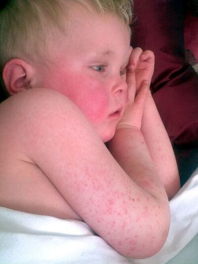
The hallmark physical finding of Scarlet Fever is the distinctive scarlet fever rash, which typically emerges within twelve to forty-eight hours following the onset of sore throat and fever. The eruption typically initiates along the upper neck, chest, and groin folds before progressing rapidly down the trunk and spreading across the peripheral extremities. Morphologically, the scarlet fever rash is composed of hundreds of minute, densely grouped erythematous maculopapules measuring one to two millimeters in diameter, elevated around hair follicles to impart an unmistakable “sandpaper” texture upon tactile palpation. When manual digital pressure is applied, the erythematous lesions completely blanch, momentarily revealing pale underlying skin before capillary refill resumes.
| Rash Parameter | Morphological Feature | Diagnostic Significance |
|---|---|---|
| Cutaneous Texture | Finely papular, pinhead-sized elevations clustered around hair follicles. |
| Imparts an unmistakable coarse sandpaper feel pathognomonic of the illness.
|
| Color and Blanching | Bright red to deep pink erythema across affected dermal zones.
| Transiently blanches completely under direct digital finger pressure.
|
| Body Distribution | Originates on neck, axillae, and groin; sweeps bilaterally over the trunk and limbs.
| Universally spares the palms and soles during the acute eruptive stage.
|
| Flexural Creases | Deep linear petechial streaks in antecubital fossae, axillae, and groin.
| Known as Pastia’s lines; caused by mechanical capillary fragility under skin folds.
|
| Facial Presentation | Intense scarlet flushing across cheeks and forehead.
| Shows striking circumoral pallor forming a pale ring around the mouth.
|
| Late Convalescence | Epidermal shedding beginning around the free borders of fingernails.
| Extensive post-exanthematous desquamation confirms prior streptococcal disease.
|
A striking diagnostic indicator of the scarlet fever rash is Pastia’s sign. In flexural regions subject to physiological skin folding and friction—most notably the antecubital fossae, axillary folds, popliteal spaces, and inguinal creases—the fragile cutaneous capillaries rupture under microvascular strain, forming confluent, deep linear petechial streaks known as Pastia’s lines. Crucially, while the cheeks and forehead display intense fiery flushing, the perioral perimeter strictly retains its natural hue, producing noticeable circumoral pallor. The eruption strictly spares the thick epidermal surfaces of the palms and soles. The scarlet fever rash persists for five to seven days before fading gradually in the order of its initial anatomical appearance. As the acute erythematous phase abates, extensive post-exanthematous desquamation begins, frequently starting as delicate flakes around the free margins of fingernails and toenails before peeling in expansive epidermal sheets across the distal extremities.
Diagnostic Recognition of the Strep Rash
In pediatric clinical triage, healthcare providers often encounter what is commonly described as a generalized strep rash. Clinicians recognize that a strep rash represents the cutaneous hypersensitivity and microvascular response triggered by group A streptococcal pyrogenic superantigens. In historical and outpatient practice, distinguishing an atypical strep rash from full-blown Scarlet Fever depends on the distribution and density of the papules. While classical Scarlet Fever presents with a diffuse, body-wide sandpaper eruption and profound systemic toxicity, a milder or focal strep rash may demonstrate patchy, localized erythema concentrated primarily around the trunk, axillae, or proximal inguinal borders.
Nevertheless, finding any sandpaper-like strep rash during an episode of documented streptococcal pharyngitis confirms that the individual strain is actively synthesizing pyrogenic exotoxins in a non-immune host. Evaluating patients with skin of color requires specific clinical diligence: erythema can be visually subtle or obscured by melanin, meaning that clinicians must rely on tactile palpation to identify follicular papularity and search meticulously for circumoral pallor, lingual papillae hypertrophy, and localized Pastia’s petechial striping. Recognizing that a subtle strep rash shares identical biological mechanisms with classic Scarlet Fever protects pediatric patients from underdiagnosis and ensures that therapeutic protocols are instituted without delay.
Diagnostic Recognition of Scarlatina Symptoms
Evaluating the comprehensive cluster of scarlatina symptoms demands systematic assessment of constitutional, mucosal, and dermal signs. The classic presentation incorporates five core physical components: high pyrexia, acute painful odynophagia, cervical lymphadenopathy, strawberry tongue changes, and a generalized scarlatiniform exanthem. Early in the course, however, scarlatina symptoms may manifest with atypical or deceptive features, particularly in children under five years of age. Toddlers and young infants frequently present without florid pharyngeal exudates, displaying instead persistent low-grade fever, irritability, mucopurulent rhinorrhea, abdominal cramping, and decreased oral fluid intake.
Understanding the full spectrum of scarlatina symptoms prevents dangerous misdiagnoses. Because the rash can be fleeting or delayed, observing tender anterior cervical lymph nodes alongside palatal petechiae and sudden-onset high fever should trigger immediate diagnostic swabbing for Scarlet Fever even prior to full cutaneous eruption. Vigilance regarding fluctuating scarlatina symptoms is essential for secondary prevention: identifying the index case halts further spread throughout classrooms and shared domestic environments.
Diagnostic Recognition of the Strep Throat Rash
Parents frequently seek urgent medical counsel when a bright strep throat rash emerges alongside their child’s complaints of severe swallowing pain. The pathophysiological link between throat pathology and the strep throat rash is mediated by the vascular absorption and systemic hematogenous circulation of streptococcal pyrogenic exotoxins from the infected tonsillar bed. The strep throat rash is fundamentally identical to the classic rash of Scarlet Fever, providing unambiguous clinical proof of a systemic streptococcal toxin state.
The sudden appearance of this sandpaper-textured strep throat rash is clinically valuable because it serves as an early warning sign that prompts immediate medical consultation, preventing patients from dismissing their sore throat as a mild viral condition. Because the strep throat rash is non-pruritic or only mildly itchy, it stands in stark contrast to urticarial allergic drug eruptions or intense contact dermatitis. Clinicians encountering a patient with an exudative tonsillopharyngitis and an emerging strep throat rash can proceed with immediate microbiological confirmation and early antimicrobial treatment for Scarlet Fever.
Clinical Diagnostic Evaluation of Scarlet Fever
Formulating an accurate diagnosis of Scarlet Fever requires synthesizing careful clinical examination with sensitive microbiological diagnostics. In outpatient primary care and emergency departments, validated clinical decision rules are frequently employed to estimate the pre-test probability of group A streptococcal infection. The Centor Score, alongside its pediatric-adapted McIsaac modification, systematically evaluates the presence of four core objective criteria: absence of cough, history of fever exceeding 38.0°C, tonsillar swelling or exudates, and tender anterior cervical lymphadenopathy. In the McIsaac scoring tool, age is incorporated as a weighting factor: patients aged three to fourteen years receive one additional point due to peak epidemiological incidence, whereas individuals aged forty-five and older lose a point due to the low baseline incidence of bacterial pharyngitis in older cohorts.
| Clinical or Demographic Criterion | Scoring Points | Clinical Guidance and Interpretation |
|---|---|---|
| Absence of Cough | +1 Point |
| Strongly favors bacterial streptococcal infection over common viral respiratory viruses.
|
| Fever History (> 38.0°C / 100.4°F) | +1 Point
| Reflects true systemic inflammatory response triggered by pyrogenic toxins.
|
| Tonsillar Swelling or Exudates | +1 Point
| Highlights localized suppurative inflammation across tonsillopharyngeal tissues.
|
| Tender Anterior Cervical Nodes | +1 Point
| Confirms regional lymphatic drainage and active immune activation against GAS.
|
| Age 3 to 14 Years | +1 Point
| Captures peak epidemiological risk group for streptococcal pharyngitis and scarlatina.
|
| Age 15 to 44 Years | 0 Points
| Represents baseline standard adult incidence profile in outpatient primary care.
|
| Age 45 Years and Older | -1 Point
| Reflects significantly reduced risk of primary streptococcal pharyngitis in older people.
|
Diagnostic Score Interpretation: Patients scoring 0 to 1 point have a low risk of streptococcal infection (<10%), meaning diagnostic swabbing and antibiotics should be withheld. A score of 2 to 3 indicates intermediate probability, warranting microbiological swab testing. A score of 4 to 5 carries a high probability (>50%), requiring a rapid antigen detection test or culture, followed by immediate antibiotic treatment upon confirmation.
While high clinical scores support strong suspicion, definitive diagnosis of Scarlet Fever requires microbiological verification. Clinicians must collect a thorough pharyngeal swab specimen by swabbing both tonsillar pillars and the posterior oropharyngeal wall vigorously, avoiding contact with the buccal mucosa, tongue, or lips. Available diagnostic modalities include:
- Rapid Antigen Detection Testing (RADT): Employs immunological methods to identify group A streptococcal cell-wall carbohydrate antigens within ten to fifteen minutes. While RADT maintains exceptional clinical specificity exceeding 95%, its clinical sensitivity varies between 70% and 90%. Consequently, international standards dictate that a negative rapid test in a symptomatic child or adolescent must be reflexively confirmed by conventional throat culture.
- Standard Throat Culture: Remains the definitive reference standard, involving inoculation onto sheep blood agar plates followed by twenty-four to forty-eight hours of aerobic and anaerobic incubation at 37°C to identify beta-hemolytic colonies sensitive to bacitracin disc diffusion.
- Molecular Nucleic Acid Amplification Tests (NAAT / PCR): Modern platforms demonstrate sensitivities and specificities approaching 100%, offering rapid automated detection that eliminates the requirement for reflex backup culture, albeit at a higher laboratory cost.
- Serological Assays (ASO and Anti-DNase B): These blood tests are completely ineffective for guiding acute antimicrobial therapy in Scarlet Fever because protective antibody titers take two to four weeks to peak. They are strictly reserved for confirming prior streptococcal exposure in patients under clinical evaluation for delayed post-infectious acute rheumatic fever or glomerulonephritis.
Diagnostic Differentials in Scarlatiniform Eruptions
Distinguishing Scarlet Fever from clinically similar childhood exanthems is essential to avoid inappropriate therapies. A critical differential diagnosis is Kawasaki disease, an acute systemic vasculitis that presents with high fever lasting at least five days, polymorphous exanthem, bilateral non-purulent conjunctivitis, anterior cervical adenopathy, mucosal erythema with a red strawberry tongue, and late desquamation of the extremities. Crucially, children with Kawasaki disease lack tonsillar exudates, fail to demonstrate an early white strawberry tongue, exhibit prominent conjunctival injection and lip fissuring, and show no clinical response to beta-lactam antimicrobials. Immediate echocardiographic assessment is mandatory in suspected Kawasaki disease to monitor for coronary artery aneurysms.
| Clinical Condition | Rash Morphology and Texture | Oral Cavity and Tongue Signs | Conjunctival Presentation | Antibiotic Response Pattern |
|---|---|---|---|---|
| Scarlet Fever | Diffuse, blanching sandpaper rash; Pastia’s lines in skin creases. |
| White to red strawberry tongue, palatal petechiae, tonsillar exudate.
| Eyes remain clear; conjunctivitis is strictly absent.
| Prompt clinical resolution occurs within 24 to 48 hours.
|
| Kawasaki Disease | Polymorphous macular rash lacking a typical gritty sandpaper texture.
| Red strawberry tongue, dry cracked fissured lips; tonsils clear.
| Bilateral non-purulent bulbar conjunctival injection is classic.
| Completely unresponsive; requires IVIG and high-dose aspirin.
|
| Measles and Adenovirus | Soft, blotchy maculopapular exanthem starting behind ears and hairline.
| Generalized pharyngeal erythema; oral Koplik’s spots in measles.
| Prominent watery conjunctivitis, marked photophobia, and coryza.
| Unresponsive to antibacterial agents; supportive viral care.
|
| Staph Scalded Skin Syndrome | Exfoliative erythema with severe tenderness and flaccid bullae.
| Normal oral mucosa; radial perioral crusting and fissuring.
| Purulent or crusting discharge; positive Nikolsky sign.
| Unresponsive to Penicillin V; requires anti-staphylococcal therapy.
|
Other potential differentials include Staphylococcal scarlet fever, which is caused by exfoliative exotoxins from Staphylococcus aureus. While Staphylococcal scarlet fever presents with a scarlatiniform sandpaper rash, it completely lacks palatal petechiae and the classic strawberry tongue, displays pronounced skin tenderness, and produces a positive Nikolsky sign. Viral exanthems, such as measles, rubella, Epstein-Barr virus infectious mononucleosis, enteroviruses, and adenoviruses, are readily differentiated by the prominence of coughing, conjunctivitis, coryza, posterior cervical or generalized adenopathy, and soft, non-gritty macular eruptions. Furthermore, widespread maculopapular drug eruptions (such as delayed hypersensitivity reactions to beta-lactams) typically present with significant cutaneous pruritus, an absence of exudative pharyngeal lesions, and a documented history of recent new medication exposure.
Evidence-Based Treatment and Clinical Practice Guidelines for Scarlet Fever
The clinical management of Scarlet Fever relies directly on prompt antimicrobial therapy, supportive symptomatic care, and patient isolation until non-infectious. Initiating appropriate treatment for Scarlet Fever accomplishes three decisive therapeutic objectives: accelerates clinical defervescence and symptom resolution, suppresses person-to-person transmission within twenty-four hours, and prevents the development of acute suppurative and non-suppurative post-streptococcal complications.
Evidence-Based Protocol: Scarlet Fever Treatment Guidelines
Modern clinical practice relies on international protocols, notably the Infectious Diseases Society of America (IDSA), the American Academy of Pediatrics (AAP), the UK Health Security Agency (UKHSA), and the European Society of Clinical Microbiology and Infectious Diseases (ESCMID), which are synthesized below in standard-of-care scarlet fever treatment guidelines. Because Streptococcus pyogenes remains universally and uniformly susceptible to beta-lactam antibiotics without documented clinical resistance anywhere in the world, penicillin and its semisynthetic derivatives remain the undisputed first-line therapy.
| Clinical Indication | Recommended Antibiotic Regimen | Dosing Schedule and Route | Treatment Duration |
|---|---|---|---|
| First-Line Standard (Oral) | Penicillin V (Phenoxymethylpenicillin) | <27 kg: 250 mg PO BID or TID; >=27 kg: 500 mg PO BID or TID. |
| 10 Full Days
|
| First-Line Pediatric (Oral) | Amoxicillin Suspension | 50 mg/kg orally once daily (max 1000 mg) or 25 mg/kg BID (max 500 mg).
| 10 Full Days
|
| Adherence / Intolerant (IM) | Benzathine Penicillin G | Single dose IM: <27 kg: 600,000 units; >=27 kg: 1,200,000 units.
| Single Dose
|
| Penicillin Allergy (Non-IgE) | Cephalexin or Cefadroxil | Cephalexin: 20 mg/kg/dose PO BID (max 500 mg/dose).
| 10 Full Days
|
| Severe Allergy (Macrolide) | Azithromycin | 12 mg/kg orally once daily (max 500 mg once daily).
| 5 Full Days
|
| Severe Allergy (Lincosamide) | Clindamycin | 7 mg/kg/dose orally three times daily (max 300 mg/dose).
| 10 Full Days
|
Under established scarlet fever treatment guidelines, the primary drug of choice is oral Phenoxymethylpenicillin (Penicillin V), prescribed for a complete ten-day course. In pediatric populations, oral Amoxicillin suspension is widely used in place of Penicillin V due to its superior palatability, reliable gastrointestinal absorption, and convenient once-daily dosing regimen (50 mg/kg once daily, maximum 1000 mg/day for 10 days). For patients with poor adherence, persistent vomiting, or challenging socio-environmental factors, a single intramuscular dose of Benzathine Penicillin G provides sustained bactericidal levels for several weeks. Adhering strictly to a full 10-day antimicrobial course remains essential to eradicate the pathogen from the pharynx and prevent acute rheumatic fever.
Targeted Antibiotic Therapy for Antibiotics Scarlet Fever
Administering targeted antibiotics scarlet fever requires careful risk stratification when selecting alternatives for beta-lactam-allergic patients. In individuals with non-immediate, mild, or non-IgE-mediated penicillin hypersensitivity (such as a delayed maculopapular rash without systemic signs), first-generation cephalosporins—such as oral cephalexin or cefadroxil for ten full days—provide safe, highly effective therapy with minimal cross-reactivity. Conversely, in patients with a history of severe Type I immediate IgE-mediated anaphylaxis, angioedema, or bronchospasm to beta-lactams, cephalosporins are contraindicated.
In severe beta-lactam allergy, physicians administering antibiotics scarlet fever utilize macrolides (oral azithromycin or clarithromycin) or lincosamides (oral clindamycin). However, widespread empirical prescribing of macrolides has led to rising resistance rates among group A streptococci internationally. Molecular surveillance programs have demonstrated that in regions such as Hong Kong, mainland China, and parts of Europe, over 60% of circulating group A streptococcal isolates associated with Scarlet Fever exhibit chromosomal or plasmid-mediated resistance to erythromycin and azithromycin. Clindamycin remains a robust alternative for beta-lactam-allergic patients and offers the added pharmacodynamic advantage of suppressing bacterial protein synthesis, directly halting the production of circulating pyrogenic exotoxins. Whenever alternative agents are employed, microbiological susceptibility confirmation is strongly advised to prevent treatment failures.
Clinical Reference Standards in Scarlet Fever Statpearls
Medical literature and clinical databases, summarized within the authoritative peer-reviewed medical repository Scarlet fever statpearls, emphasize several core clinical principles:
- Prompt Treatment: Emphasizes that antibiotic therapy must be initiated as soon as group A streptococcal infection is confirmed, while reinforcing that initiating treatment within nine days of acute symptom onset remains fully protective against acute rheumatic fever.
- Contagion Mitigation: Reaffirms that patients cease to be infectious and may safely return to educational, childcare, or occupational settings precisely twenty-four hours after the first therapeutic dose of antibiotics, provided that pyrexia has resolved.
- Avoidance of Post-Treatment Swabs: Recommends against performing routine follow-up throat cultures or rapid testing in asymptomatic patients following completion of therapy. Approximately 10% to 25% of treated individuals become chronic, asymptomatic pharyngeal group A streptococcal carriers who face no risk of complications and do not spread disease.
- Supportive Regimens: Emphasizes comprehensive supportive care, including weight-based acetaminophen (paracetamol) or ibuprofen for pyrexia and odynophagia, vigorous oral fluid rehydration, cool humidification, and avoiding aspirin in all individuals younger than nineteen years of age to prevent Reye’s syndrome.
Suppurative and Non-Suppurative Complications of Scarlet Fever
The clinical gravity of Scarlet Fever is underscored by its capacity to produce both immediate suppurative infections through local tissue invasion and delayed non-suppurative autoimmune sequelae mediated by molecular mimicry. Timely antimicrobial administration radically reduces these complications.
| Pathological Category | Complication Manifestation | Pathophysiological Mechanism | Clinical Presentation and Impact |
|---|---|---|---|
| Suppurative (Local) | Peritonsillar Abscess (Quinsy) | Direct bacterial invasion into peritonsillar space. |
| Severe odynophagia, hot-potato voice, uvular deviation, trismus.
|
| Suppurative (Local) | Retropharyngeal Abscess | Spread into posterior pharyngeal fascial spaces.
| Stridor, neck stiffness, toxic appearance, airway compromise.
|
| Suppurative (Regional) | Otitis Media & Mastoiditis | Direct extension along the Eustachian tube.
| Severe ear pain, tympanic rupture, temporal tenderness.
|
| Suppurative (Invasive) | Invasive Strep & Sepsis (iGAS) | Hematogenous spread to deep sterile body sites.
| Bacteremia, necrotizing fasciitis, meningitis, pneumonia.
|
| Suppurative (Systemic) | Toxic Shock Syndrome (STSS) | Massive cytokine release driven by superantigens.
| Sudden hypotension, multi-organ failure; 30% to 50% mortality.
|
| Non-Suppurative (Cardiac) | Acute Rheumatic Fever (ARF) | Molecular mimicry against human cardiac myosin.
| Migratory arthritis, carditis, chronic mitral valve disease.
|
| Non-Suppurative (Renal) | Post-Strep Glomerulonephritis | Deposition of circulating immune complexes in glomeruli.
| Hematuria (tea-colored urine), edema, hypertension, oliguria.
|
| Non-Suppurative (Joint) | Post-Strep Reactive Arthritis | Immune complex-mediated synovial inflammation.
| Non-migratory peripheral joint swelling within 10 days.
|
| Non-Suppurative (Neural) | PANDAS Syndrome | Autoantibodies binding to neuronal basal ganglia.
| Rapid-onset obsessive-compulsive tics and behavioral changes.
|
Suppurative Complications
These occur when Streptococcus pyogenes spreads directly into adjacent structures or through regional lymphatic and hematogenous routes.
- Peritonsillar and Retropharyngeal Abscesses: Manifest with severe localized throat pain, persistent high fever, hot-potato voice, trismus, and unilateral deviation of the uvula, necessitating immediate parenteral antibiotics and surgical needle aspiration or drainage.
- Acute Otitis Media and Mastoiditis: Result from ascension of bacteria along the Eustachian tube into the middle ear and temporal bone air cells.
- Invasive Group A Streptococcal (iGAS) Disease: Occurs when bacteria enter deeper anatomical structures, causing necrotizing soft tissue infections, pneumonia, purulent meningitis, endocarditis, or bacteremic sepsis.
- Streptococcal Toxic Shock Syndrome (STSS): Severe superantigen-mediated systemic collapse characterized by acute hypotension, capillary leak syndrome, and rapid multi-organ failure, carrying mortality rates of 30% to 50%.
Non-Suppurative Autoimmune Sequelae
These develop when cross-reactive host antibodies damage native tissues via molecular mimicry.
- Acute Rheumatic Fever (ARF): Develops two to four weeks after an untreated pharyngeal group A streptococcal infection. It presents with migratory polyarthritis, carditis, Sydenham’s chorea, erythema marginatum, and subcutaneous nodules (evaluated using the revised Jones criteria). Persistent autoimmune valvular injury leads to chronic rheumatic heart disease, most frequently manifesting as mitral valve stenosis. Antibiotic therapy initiated within nine days of symptom onset prevents ARF.
- Post-Streptococcal Glomerulonephritis (PSGN): Arises one to three weeks following pharyngeal or cutaneous streptococcal infection. Immune complex deposition within renal glomerular basement membranes activates complement pathways, producing acute nephritic syndrome characterized by gross hematuria (“tea-colored” urine), periorbital and peripheral edema, hypertension, and acute kidney injury. Unlike rheumatic fever, the development of PSGN is not reliably prevented by antibiotic therapy.
- Post-Streptococcal Reactive Arthritis (PSRA): Causes non-migratory joint inflammation that develops within ten days of infection and does not follow the classic course of acute rheumatic fever.
- PANDAS: Pediatric Autoimmune Neuropsychiatric Disorders Associated with Streptococcal Infections describes an acute exacerbation of obsessive-compulsive symptoms or motor/vocal tics following group A streptococcal exposure, driven by autoantibodies targeting basal ganglia neurons.
Epidemiology, Transmission, and Public Health Prevention of Scarlet Fever
Scarlet Fever is a human-specific infection; Streptococcus pyogenes has no recognized animal reservoir. Transmission occurs primarily through large respiratory droplets expelled during coughing, sneezing, or close interpersonal conversation. Direct physical contact with contaminated secretions, unpasteurized milk, or contaminated food can also trigger localized foodborne outbreaks. Secondary household attack rates are high, with close contacts facing an estimated 35% probability of acquiring the pathogen. The incubation period ranges from two to five days.
| Transmission Vector | Mechanism of Exposure | Target Preventive Measure |
|---|---|---|
| Airborne Droplets | Inhalation of large aerosols from coughing or sneezing. |
| Cover mouth and nose with tissues; discard immediately.
|
| Direct Contact | Touching skin lesions or infected oral secretions.
| Frequent handwashing for 20 seconds with soap and water.
|
| Indirect Fomites | Handling shared drinking glasses, cutlery, or towels.
| Prohibit sharing personal items; wash in hot soapy water.
|
| Contaminated Food | Ingestion of unpasteurized milk or tainted food items.
| Enforce strict commercial pasteurization and food hygiene.
|
Epidemiologically, Scarlet Fever predominantly affects children between three and eight years of age, with peak incidence spanning five to fifteen years; it remains rare in infants under twelve months due to protective maternal antibodies. In temperate zones, infection follows a seasonal rhythm, peaking during late autumn, winter, and early spring. Global surveillance reveals significant regional shifts. Starting in 2011, major resurgence waves were recorded across East Asia, including Hong Kong, mainland China, and South Korea, where the national incidence in South Korea increased from 0.3 per 100,000 in 2008 to 13.7 per 100,000 in 2015, dominated by emm4, emm28, emm1, and emm3 lineages alongside rising macrolide and tetracycline resistance. Similarly, the United Kingdom documented extensive surges between 2014 and the 2022–2023 season, marked by a 68% rise in notifications and parallel increases in invasive group A streptococcal (iGAS) infections.
Infection Control and School Exclusion Criteria
Because there is currently no licensed human vaccine against Streptococcus pyogenes, prevention relies entirely on non-pharmacological infection control and prompt antibiotic stewardship. The vast diversity of M protein types (>220 emm variants) has historically complicated vaccine development, although modern multivalent and conserved-region recombinant vaccine candidates remain under clinical investigation. Public health regulations mandate that Scarlet Fever is a statutory notifiable disease in numerous jurisdictions, including the United Kingdom, China, and South Korea.
Effective community containment protocols require:
- Strict Isolation and Exclusion: Children diagnosed with Scarlet Fever must remain strictly excluded from schools, childcare centers, and sports activities until twenty-four hours after starting their prescribed antibiotic regimen, provided their fever has fully subsided. Untreated individuals remain highly infectious for two to three weeks.
- Hand and Environmental Hygiene: Frequent handwashing with liquid soap and warm water for at least twenty seconds, or use of 70% to 80% alcohol-based sanitizers. Contaminated environmental surfaces, toys, and utensils should be disinfected using a 1:99 dilution of household bleach.
- Respiratory Etiquette: Use of disposable tissues to cover the mouth and nose during coughing or sneezing, followed by immediate disposal in covered receptacles and hand cleaning. Avoid sharing drinking bottles, cups, plates, or linens within households.
Frequently Asked Questions
What is scarlet fever?
Scarlet Fever is an acute, contagious bacterial infection caused by Streptococcus pyogenes, also known as Group A Streptococcus. It most commonly develops in association with streptococcal pharyngitis (strep throat) when the infecting bacterial strain secretes specific pyrogenic exotoxins that enter the systemic circulation. The disease is clinically characterized by the sudden onset of a high fever, a painful sore throat, a distinctive bright red “strawberry tongue,” and a widespread red sandpaper-like rash. Although historically dangerous, it is today fully treatable with a standard course of antibiotics prescribed by a healthcare provider.
Is scarlet fever contagious?
Yes, Scarlet Fever is highly contagious and spreads easily from person to person through respiratory secretions and airborne droplets. When an infected individual coughs, sneezes, speaks, or breathes, infectious droplets can be inhaled by nearby individuals or transferred to surfaces and shared utensils. A person with untreated Scarlet Fever can remain infectious for two to three weeks after the initial onset of symptoms. However, once a patient completes the first twenty-four hours of appropriate antibiotic treatment, they are no longer contagious and may safely return to school or work.
Is scarlet fever dangerous?
While Scarlet Fever was once a severe, life-threatening childhood illness with high mortality, it is rarely dangerous today when treated promptly with antibiotics. However, if the infection is ignored or left untreated, the bacteria can spread locally and systemically to cause severe suppurative conditions like peritonsillar abscesses, ear infections, pneumonia, or life-threatening streptococcal toxic shock syndrome. Furthermore, untreated cases can trigger delayed immune-mediated complications, most notably acute rheumatic fever—which can permanently damage heart valves—and acute post-streptococcal kidney inflammation. Seeking early clinical evaluation and completing all prescribed medications keeps the risk of severe complications exceptionally low.
Is the scarlet fever rash contagious?
No, the scarlet fever rash itself is not directly contagious, and a person cannot contract the disease simply by coming into physical contact with the reddened skin or peeling epidermal flakes. The cutaneous eruption is not caused by active live bacteria living in the superficial layers of the skin, but is instead an inflammatory and hypersensitivity reaction caused by circulating bacterial toxins interacting with blood vessels. However, the person carrying the rash is actively contagious through their nose, throat, and oral respiratory fluids. Therefore, precautions should focus on preventing the transmission of respiratory droplets and mouth secretions rather than avoiding touch with the rash.
How serious is scarlet fever?
In modern medical settings, Scarlet Fever is generally classified as a mild-to-moderate illness that responds rapidly to standard antibiotic regimens. Most children experience noticeable relief, fever resolution, and fading of the exanthem within twenty-four to forty-eight hours of beginning antibiotic therapy. Nevertheless, Scarlet Fever must always be treated with medical seriousness because failure to complete the full course of antibiotics can lead to recurrent infection and late complications. Severe outcomes such as rheumatic heart disease and invasive bacterial spread illustrate why medical guidelines uniformly mandate a full ten-day course of penicillin or an approved equivalent.
The following posts may interest you
Fifth Disease (slapped cheek syndrome): Symptoms, Risks & Care
Sixth Disease (Roseola): Symptoms, Rash, and Treatment
When Should I Call the Doctor for a Fever in My Baby?
Sources
Pharyngitis and Scarlet Fever
https://www.ncbi.nlm.nih.gov/books/NBK333418
Incidence and Characteristics of Scarlet Fever, South Korea, 2008–2015
https://pmc.ncbi.nlm.nih.gov/articles/PMC5367408
The incidence of scarlet fever
The rash with maculopapules and fever in children
https://www.sciencedirect.com/science/article/abs/pii/S0738081X18302530
Incidence of Scarlet Fever in Children in Jeju Province, Korea, 2002-2016: An Age-period-cohort Analysis
https://pmc.ncbi.nlm.nih.gov/articles/PMC6549015
THE HISTORY OF SCARLET FEVER
https://pmc.ncbi.nlm.nih.gov/articles/PMC2456687
Patterns in the Incidence of Scarlet Fever Among Children Aged 0–9 Years — China, 2010–2019
https://pmc.ncbi.nlm.nih.gov/articles/PMC10485360
Epidemiology of Reemerging Scarlet Fever, Hong Kong, 2005–2015
https://pmc.ncbi.nlm.nih.gov/articles/PMC5621532
Scarlet Fever Outbreak, Hong Kong, 2011
https://pmc.ncbi.nlm.nih.gov/articles/PMC3471616
Some Factors Influencing the Spread of Scarlet Fever in an Institution
image: By www.badobadop.co.uk – Own work, CC BY-SA 3.0, https://commons.wikimedia.org/w/index.php?curid=30524904