Parainfluenza: Key Facts at a Glance
Parainfluenza is a group of four common respiratory viruses (HPIV-1 to HPIV-4). Despite the name, it is not the flu. In healthy adults and older children it usually causes cold-like symptoms. In babies and young children it is a leading cause of croup, bronchiolitis, and viral pneumonia. It is most serious in infants under 6 months and in people with weakened immune systems.
- Symptoms: A runny nose, sore throat, sneezing, low-grade fever, and cough. In young children it often causes croup: a seal-like barking cough, a hoarse voice, and noisy breathing (stridor) that gets worse at night.
- How it spreads: Through droplets from coughs, sneezes, and talking at close range, and by touching contaminated surfaces such as toys, tables, and doorknobs and then the eyes, nose, or mouth. The virus can survive on hard surfaces for up to 10 hours.
- Timeline: Symptoms begin 2 to 6 days after exposure. Mild illness and mild croup usually clear up within 7 to 10 days with care at home, but a cough can linger for 2 to 4 weeks.
- Treatment: There is no approved antiviral medicine. Care is supportive: fluids, rest, a cool-mist vaporizer, and acetaminophen or ibuprofen for fever. Do not give aspirin to children. Antibiotics do not work against the virus. Doctors treat severe croup with dexamethasone and nebulized epinephrine.
- Prevention: There is no licensed vaccine. Wash hands with soap and water for 20 seconds, clean high-touch surfaces, cover coughs and sneezes, and keep sick children home until the fever is gone.
- When to get emergency care: Trouble breathing, fast or labored breathing, the chest pulling in with each breath, noisy breathing at rest, pale or bluish lips, extreme sleepiness, confusion, or being unable to drink. In babies, poor feeding with far fewer wet diapers needs urgent medical attention.
Understanding Parainfluenza: Virology, Pathology, and Clinical Syndromes
Parainfluenza viruses are ubiquitous enveloped pathogens that establish clinically diverse respiratory infections across pediatric, adult, and immunocompromised patient cohorts. Belonging to the family Paramyxoviridae, the Parainfluenza group represents a substantial medical burden, serving as the second leading viral cause of hospital admissions for pediatric lower respiratory illnesses globally. Because Parainfluenza exhibits diverse structural phenotypes and antigenic profiles, the induced manifestations range from mild rhinorrhea and pharyngitis to life-threatening subglottic inflammation and acute alveolar injury. A thorough scientific assessment of Parainfluenza requires investigating viral genomics, virion ultrastructure, immune evasion, seasonal epidemiology, differential diagnosis, and current clinical guidelines.
The physiological impact of Parainfluenza within clinical environments extends from simple self-limiting coryza to protracted intensive care admissions. Primary infection with Parainfluenza occurs very early in childhood, establishing a recurrent pattern of reinfections that persist throughout adult life due to incomplete long-term mucosal immunity. By dissecting the immunological characteristics and molecular behaviors of Parainfluenza, healthcare professionals can implement targeted clinical triage, avoid the inappropriate administration of antibacterial agents, and deliver optimal supportive care.
Virology Basics and Structural Biology of Parainfluenza
The architectural integrity of the Parainfluenza virion relies on a pleomorphic lipid bilayer envelope encasing a negative-sense, single-stranded ribonucleic acid (vRNA) genome. The genome of Parainfluenza spans approximately 15,000 nucleotides organized in a linear sequence that encodes essential structural and nonstructural proteins. Efficient replication and transcription adhere strictly to the classic “rule of six,” a genomic feature wherein the total nucleotide length must remain divisible by six for optimal encapsidation and enzymatic fidelity.
| Structural Component | Molecular Weight (kDa) | Biological Function in Infection |
|---|---|---|
| Nucleocapsid Protein (NP/N) | 66–70 | Coats genomic RNA in a 1:6 ratio, creating the functional ribonucleoprotein core template. |
| Phosphoprotein (P) | 49–90 | Functions as a vital cofactor facilitating RNA-dependent RNA polymerase binding and transcription. |
| Large Polymerase (L) | 175–251 | Catalyzes negative-strand transcription, mRNA capping, polyadenylation, and genomic replication. |
| Matrix Protein (M) | 28–40 | Stabilizes virion geometry beneath the inner lipid leaflet and drives budding dynamics. |
| Fusion Glycoprotein (F) | 60–66 | Mediates cellular entry and host-cell membrane coalescence following endoproteolytic cleavage. |
| Hemagglutinin-Neuraminidase (HN) | 69–82 | Directs receptor attachment to terminal sialic acids and enzymatically facilitates newly formed virion release. |
Transcription commences when the Parainfluenza polymerase complex accesses the unsegmented ribonucleoprotein. Beyond structural duties, nonstructural proteins—such as the C, V, and D accessory products—act as potent immunological shields by degrading STAT proteins and suppressing interferon cascades, allowing Parainfluenza to establish persistent cellular colonization.
Taxonomic Classification and Characteristics of Parainfluenza Serotypes
Taxonomically, human Parainfluenza pathogens diverge into two distinct genera within the subfamily Paramyxovirinae. The genus Respirovirus includes human parainfluenza virus type 1 and human parainfluenza 3, whereas the genus Rubulavirus encompasses human parainfluenza 2 and human parainfluenza 4 (subtypes 4A and 4B). Understanding this fundamental genetic taxonomy clarifies the notable variability in pathogenicity, age distribution, and clinical progression seen across Parainfluenza outbreaks.
Clinical Taxonomy Insight:
While the umbrella label piv virus is frequently used in bedside settings, each serotype occupies a distinct pathophysiological and anatomical niche within the respiratory system.
Pathophysiological Divergence of Human Parainfluenza Serotypes
- Respirovirus Dynamics: Both human parainfluenza virus type 1 and human parainfluenza 3 possess heavier phosphoproteins and exhibit heightened tropism for human respiratory tract epithelia. Human parainfluenza 3 binds broader terminal sialic acid configurations, explaining its ability to penetrate deeper into distal bronchial passages.
- Rubulavirus Dynamics: Human parainfluenza 2 and human parainfluenza 4 synthesize distinctive accessory products like the cysteine-rich V protein, which slows host mitotic activity and blocks interferon-alpha and beta signaling.
Transmission Dynamics, Environmental Stability, and Pathogenesis of Parainfluenza
The primary transmission modality for Parainfluenza involves large respiratory droplets expelled during coughing, sneezing, or verbalization. Unlike smaller aerosolized virions, Parainfluenza relies heavily on close physical proximity and fomite contamination. Virions exhibit pronounced survival on hard nonporous surfaces, remaining infectious for up to ten hours, and persist on porous fabrics for four hours. In contrast, personal cutaneous transfer via fingertip inoculation degrades rapidly within ten minutes.
| Stage of Pathogenesis | Physiological Mechanism | Anatomical Progression & Tissue Effects |
|---|---|---|
| Phase 1: Inoculation | Inhalation of infectious respiratory droplets or direct digital contact with contaminated fomites. | Viral particles lodge on the pseudostratified ciliated epithelium of the nose and throat. |
| Phase 2: Viral Attachment | Hemagglutinin-neuraminidase (HN) spikes selectively bind terminal sialic acid residues on epithelial surfaces. | Stable attachment to host cell membranes occurs without immediate host immune activation. |
| Phase 3: Proteolytic Entry | Host proteases (such as furin) cleave the precursor F0 glycoprotein into active F1 and F2 subunits. | Direct viral-to-host membrane fusion releases the ribonucleoprotein core into the cytoplasm. |
| Phase 4: Cellular Injury | Viral transcription and translation trigger ciliary arrest, focal rounding, and syncytia formation. | Mucosal desquamation, focal inflammatory infiltration, and subglottic or bronchial narrowing. |
Pathological Note on Tissue Destruction:
The epithelial damage inflicted by Parainfluenza typically induces transient ciliary clearance arrest, leaving airway structures vulnerable to secondary bacterial colonization while inflammatory mononuclear infiltrates gather along mucosal basolateral zones.
Pathogenesis begins within the pseudostratified ciliated columnar cells of the nasopharynx. Parainfluenza attaches via viral hemagglutinin-neuraminidase spikes to cell surface sialic acid receptors. Cellular penetration occurs when host endoproteases cleave the inactive F0 glycoprotein into active disulfide-linked F1 and F2 subunits. As Parainfluenza replicates, host mitotic activity declines, triggering cytoplasmic vacuolization, basophilic inclusion bodies, and multinucleated syncytia formation.
Epidemiological Surveillance and Seasonality of Parainfluenza
Surveillance records from national monitoring programs demonstrate distinct cyclical trends among Parainfluenza serotypes. While Parainfluenza persists at baseline levels throughout every calendar month, predictable seasonal spikes define regional public health burdens.
| Serotype Designation | Epidemic Pattern | Peak Calendar Occurrence | Primary Vulnerable Population |
|---|---|---|---|
| HPIV-1 | Biennial cycles | Autumn months of odd-numbered years | Toddlers aged 7 to 36 months |
| HPIV-2 | Biennial or annual cycles | Autumn to early winter of even-numbered years | Children aged 1 to 2 years |
| HPIV-3 | Annual endemic peaks | Spring and early summer | Infants younger than 6 months |
| HPIV-4 | Annual endemic peaks | Fall through winter months | Young children, school-age youth, adults |
The emergence of human parainfluenza 3 in spring frequently anticipates lower airway disease prior to winter outbreaks of influenza and respiratory syncytial virus. Furthermore, coinfections involving Parainfluenza alongside rhinoviruses or adenoviruses occur in nearly a third of diagnosed pediatric cases, compounding clinical complexity.
Comprehensive Clinical Manifestations Across Patient Groups
The clinical spectrum of Parainfluenza reflects a continuum spanning upper and lower respiratory structures. Following an incubation phase lasting two to six days, infected patients present with initial rhinorrhea, low-grade pyrexia, sneezing, and pharyngeal irritation. The downstream impact of Parainfluenza hinges on anatomical vulnerability, baseline pulmonary reserve, and patient immunological competence.
Classical Upper Airway Syndromes
- Acute Laryngotracheobronchitis (Croup): The signature manifestation of pediatric Parainfluenza involves extensive subglottic laryngeal edema. Because the non-distensible cricoid cartilage rings the upper trachea, focal inflammation quickly precipitates luminal narrowing. Children experience a hallmark seal-like barking cough, hoarseness, and inspiratory stridor that intensifies during nocturnal hours.
- Tracheobronchitis: In school-age children and immunocompetent adults, Parainfluenza frequently manifests as an aggressive tracheobronchitis characterized by deep productive coughing, substernal tightness, and coarse rhonchi on auscultation.
Classical Lower Airway Syndromes
- Acute Bronchiolitis: When Parainfluenza spreads into terminal bronchioles, patients experience profound air trapping, tachypnea, expiratory wheezing, subcostal retractions, and scattered rales. Young infants under six months are at high risk due to their minimal baseline airway caliber.
- Viral and Secondary Bacterial Pneumonia: Parainfluenza triggers parenchymal lung consolidation, diffuse interstitial infiltrates, and severe alveolar-capillary gas exchange defects. Bacterial superinfections involving organisms such as Streptococcus pneumoniae or Staphylococcus aureus can emerge following mucosal injury.
Unique Clinical Syndromes and At-Risk Populations
The severity of Parainfluenza escalates dramatically in distinct vulnerable populations. Pediatric patients exhibit anatomical susceptibilities, whereas geriatric and immunocompromised individuals face severe systemic and pulmonary sequelae.
Pediatric Manifestations: Parainfluenza symptoms in kids
Primary infection with Parainfluenza typically occurs early in childhood. Classic Parainfluenza symptoms in kids include sudden fever, nasal blockage, barking cough, and respiratory fatigue. Young infants infected with human parainfluenza 3 can develop severe feeding intolerance, lethargy, cyanosis, and dehydration secondary to rapid breathing. Furthermore, acute otitis media occurs in up to 60% of pediatric viral respiratory episodes, with Parainfluenza frequently identified in middle ear aspirates.
Immunocompromised Hosts and Hematopoietic Stem Cell Recipients
In adult or pediatric bone marrow transplant recipients and patients with severe combined immunodeficiency, Parainfluenza can cause destructive giant-cell pneumonia. Unlike immunocompetent hosts, who clear the virus in one to two weeks, immunocompromised patients may continuously shed viable Parainfluenza virions for several months. Reported mortality ranges between 22% and 75% once lower tract parenchymal consolidation is established.
Extrapulmonary Dissemination and Rare Complications
Although Parainfluenza remains predominantly restricted to respiratory surfaces, hematogenous viremia can occasionally deliver viral components into distant tissues. Reported extrapulmonary presentations include:
- Neurological Infiltration: Febrile convulsions occur frequently in pediatric Parainfluenza infections. On rare occasions, viral antigens have been isolated from cerebrospinal fluid in cases of meningoencephalitis, ventriculitis, and Guillain-Barré syndrome.
- Myopericarditis and Parotitis: Direct parenchymal inflammation of salivary glands mimicking mumps, as well as acute myocarditis and rhabdomyolysis, have been documented in isolated Parainfluenza clinical reports.
Etiology: Comprehensive Pathogen Analysis
A rigorous clinical assessment of Parainfluenza requires categorizing primary and secondary factors that shape viral proliferation, host vulnerability, and symptom progression.
| Etiological Dimension | Primary Factors Involved | Pathophysiological Impact on Disease Severity |
|---|---|---|
| Primary Viral Agents | HPIV-1, HPIV-2, HPIV-3, HPIV-4 serotypes (Paramyxoviridae). | Direct cytotoxic destruction of airway epithelium via viral surface glycoproteins. |
| Host Anatomy | Pediatric subglottic cricoid ring diameter and small airway caliber. | Minor mucosal swelling produces exponential resistance to laminar airflow. |
| Immune Vulnerability | Immature cellular immunity, waning maternal antibodies, organ transplantation. | Inability to limit viral replication, promoting chronic viral shedding and giant-cell formation. |
| Environmental Triggers | Passive smoke inhalation, industrial air pollutants, overcrowding. | Compromises mucociliary escalator defense, increasing viral attachment rates. |
Primary Viral Etiology: Causes
The direct biological trigger of Parainfluenza illness is mucosal infection by one of the four human paramyxovirus serotypes. Viral membrane glycoproteins drive target attachment and tissue ingress. Each subtype possesses a unique tropism:
- HPIV-1: Primary viral cause of laryngotracheobronchitis and subglottic swelling.
- HPIV-2: Secondary cause of croup and upper respiratory inflammatory syndromes.
- HPIV-3: Leading cause of bronchiolitis and interstitial pulmonary involvement in infants.
- HPIV-4: Etiological agent behind mild-to-moderate tracheobronchitis and cold syndromes.
Secondary Contributing Factors: Causes
Several physiological, immunological, and environmental elements aggravate Parainfluenza progression:
- Immunological Immaturity: Waning maternal transplacental IgG antibodies around six months of age exposes infants to severe primary Parainfluenza infection.
- Environmental Toxin Exposure: Inhaled tobacco smoke and ambient particulate pollutants impair mucociliary escalator clearance, increasing viral entry.
- Nutritional Deprivation: Vitamin A deficiency and childhood protein malnutrition directly disrupt epithelial mucosal repair mechanisms.
- Anatomical Subglottic Dimensions: Pediatric laryngeal architecture is easily compromised by minor amounts of inflammatory swelling.
Comprehensive Clinical Symptom Profiles: Symptoms
Recognizing the complex symptom array of Parainfluenza enables timely clinical triage and prevents unnecessary diagnostic delays.
| Symptom Category | Pathophysiological Mechanism | Clinical Manifestation |
|---|---|---|
| Systemic Pyrexia | Cytokine release (IL-1, IL-6, TNF-alpha) from stimulated immune cells. | Low-grade fever in adults, spikes up to 40°C in pediatric croup. |
| Upper Airway Obstruction | Subglottic mucosal edema and vocal cord inflammatory infiltration. | Harsh, seal-like barking cough, hoarseness, inspiratory stridor. |
| Bronchial Irritation | Ciliary sloughing, hyperactive sensory neuropeptide stimulation. | Dry-to-productive cough, chest discomfort, wheezing. |
| Peripheral Alveolar Failure | Alveolar exudate, air trapping, interstitial thickening. | Tachypnea, nasal flaring, intercostal retractions, cyanosis. |
Early Clinical Indications: Symptoms
Initial Parainfluenza manifestations closely mirror typical upper respiratory tract viral infections:
- Clear-to-mucoid anterior nasal discharge
- Pharyngeal hyperaemia and scratchy throat discomfort
- Mild ocular conjunctival injection and sneezing
- Generalized fatigue, malaise, and frontal headaches
Progressive Severe Indications: Symptoms
When infection extends past the larynx into pulmonary zones, Parainfluenza displays critical signs of respiratory compromise:
- Persistent biphasic or resting inspiratory stridor
- Subcostal and sternocleidomastoid muscle retractions
- Decreased oral fluid intake and signs of systemic dehydration
- Lethargy, progressive somnolence, confusion, or peripheral cyanosis
Diagnostic Modalities and Clinical Laboratory Evaluation
Accurate differentiation of Parainfluenza from other respiratory pathogens requires combining clinical evaluation with laboratory-confirmed molecular diagnostics. Because symptoms overlap significantly with respiratory syncytial virus, adenovirus, and influenza, empirical identification alone is frequently inconclusive.
| Diagnostic Step | Clinical Action Required | Key Diagnostic Value |
|---|---|---|
| Step 1: Clinical Triage | Assess work of breathing, resting stridor, and air entry. | Establishes immediate severity score and urgency of airway stabilization. |
| Step 2: Swab Acquisition | Collect deep nasopharyngeal secretions using flocked synthetic swabs. | Maximizes intact epithelial cell harvest for molecular and antigen assays. |
| Step 3: Molecular Assay | Perform multiplex reverse-transcription polymerase chain reaction (RT-PCR). | Rapid, definitive detection of specific serotypes (HPIV-1 to HPIV-4) within hours. |
| Step 4: Supportive Imaging | Obtain soft-tissue neck and chest radiographs when indicated. | Visualizes the subglottic steeple sign in croup or interstitial pulmonary infiltrates. |
Diagnostic Gold Standard Shift:
While traditional viral culture in monkey kidney cells was historically deemed the diagnostic standard, modern multiplex RT-PCR testing provides significantly higher sensitivity, specificity, and rapid turnaround required for immediate infection control decisions.
Specimen Collection and Pre-Analytical Considerations
The concentration of shed Parainfluenza virions peaks within the first two to four days of symptomatic illness. Optimal diagnostic yields depend on using synthetic flocked swabs stored in standard viral transport media buffered to physiological pH. Nasopharyngeal aspirates and nasal washes provide superior sensitivity in infants compared to simple dry swabs. For lower tract evaluations in mechanically ventilated patients, bronchoalveolar lavage fluid or deep endotracheal secretions should be tested.
Diagnostic Modalities Compared
- Reverse-Transcription Polymerase Chain Reaction (RT-PCR): Multiplex molecular panels serve as the contemporary gold standard for Parainfluenza detection. Commercial platforms identify individual serotypes within a matter of hours, delivering sensitivities and specificities exceeding 95%.
- Direct Immunofluorescence Assays (DFA): Utilizing conjugated monoclonal antibodies, direct immunofluorescence visualizes viral antigens directly within exfoliated epithelial cells. Sensitivities range from 65% to 85%.
- Conventional Viral Isolation Culture: Historically regarded as the standard, primary rhesus or cynomolgus monkey kidney cell lines facilitate Parainfluenza propagation. Confirmation traditionally relies on guinea pig erythrocyte hemadsorption, though turnaround times require several days to three weeks.
- Serological Antibody Panels: Paired acute and convalescent sera assessing a four-fold increase in neutralizing or hemagglutinin-inhibition antibodies are historically informative but hold minimal acute clinical utility due to widespread heterotypic cross-reactivity.
- Radiological Imaging: In pediatric croup, anteroposterior soft-tissue neck radiographs may reveal symmetrical subglottic narrowing known as the “steeple sign,” while chest imaging in lower tract disease demonstrates peribronchial cuffing, hyperinflation, or patchy consolidation.
Therapeutic Interventions and Patient Management: Treatment
Presently, no targeted antiviral medications have received regulatory approval for treating Parainfluenza infections. Consequently, therapeutic management focuses on supportive care, airway maintenance, and addressing anatomical obstructions.
| Care Setting | Core Therapeutic Protocol | Clinical Objectives & Mechanism |
|---|---|---|
| Outpatient Home Care | Oral hydration, cool-mist vaporizers, and antipyretics (acetaminophen/ibuprofen). | Maintains systemic fluid balance, relieves pharyngeal irritation, and controls pyrexia. |
| Emergency Laryngeal Triage | Nebulized racemic epinephrine along with systemic dexamethasone (0.6 mg/kg). | Induces rapid mucosal alpha-adrenergic vasoconstriction while reducing laryngeal edema. |
| Inpatient Hospital Ward | Humidified supplemental oxygen, intravenous fluids, and continuous pulse oximetry. | Stabilizes peripheral oxygen saturation above 92% and mitigates metabolic exhaustion. |
| Immunocompromised Specialty Care | Experimental investigational agents (inhaled DAS181 or aerosolized ribavirin). | Blocks viral sialic acid binding sites or disrupts paramyxoviral polymerase activity. |
Antibiotic Stewardship Caution:
Because Parainfluenza is fundamentally an acute viral syndrome, antimicrobial medications offer zero efficacy and must not be prescribed unless secondary bacterial superinfections are unequivocally verified.
General Supportive Care Interventions: Treatment
For most immunocompetent patients experiencing mild upper respiratory forms of Parainfluenza, conservative outpatient care suffices:
- Systemic Hydration: Maintaining adequate oral fluid volume thins inspissated mucosal secretions and prevents dehydration.
- Airway Humidification: Cool-mist vaporizers or warm bathroom steam assist in loosening dried upper airway exudates.
- Antipyretic Administration: Acetaminophen and ibuprofen relieve fever and bodily discomfort. Aspirin is strictly contraindicated in pediatric cohorts due to the risk of Reye’s syndrome.
- Rest and Calming Measures: Minimizing physical agitation decreases negative inspiratory pressures, stabilizing airway flow in struggling toddlers.
Pharmacological and Hospital-Based Interventions: Treatment
When Parainfluenza causes acute respiratory compromise or severe croup, escalated medical treatment is vital:
- Corticosteroid Therapy: Systemic dexamethasone (administered as a single oral or intramuscular dose of 0.15 to 0.6 mg/kg) acts as a cornerstone therapy, mitigating subglottic edema within two to four hours.
- Nebulized Epinephrine: In moderate-to-severe stridor at rest, nebulized racemic or L-epinephrine produces local mucosal alpha-adrenergic vasoconstriction, providing rapid symptomatic relief within 30 minutes.
- Oxygen Supplementation: Humidified oxygen delivered through nasal cannulas or high-flow circuits maintains arterial oxygen saturation above 92%.
- Avoidance of Antibacterial Misuse: Because Parainfluenza is a viral illness, standard antibacterial drugs are ineffective and should be withheld unless secondary bacterial tracheitis or pneumonia is confirmed.
Investigational Antiviral Strategies: Treatment
In immunocompromised individuals suffering from invasive lower tract Parainfluenza, several therapeutic candidates continue to undergo clinical evaluation:
- DAS181: A recombinant inhaled host-directed sialidase that cleaves terminal sialic acid receptors from respiratory epithelial linings, preventing virion binding and cell entry.
- Aerosolized Ribavirin: Investigated in stem cell transplant recipients, though data demonstrating broad survival benefits remain limited.
- Fusion-Inhibitory Peptides: Engineered heptad-repeat synthetic peptide polymers targeting the F glycoprotein core have demonstrated promising viral suppression in laboratory models.
Preventive Protocols and Infection Control Strategies: Prevention
Because licensed preventative vaccines for Parainfluenza are not currently available, infection control relies on non-pharmaceutical hygiene measures and environmental barriers. Preventing transmission is critical in pediatric wards, neonatal units, and oncology clinics.
| Prevention Strategy | Underlying Mechanism | Clinical Implementation |
|---|---|---|
| Meticulous Hand Hygiene | Dissolves viral lipid envelope via detergents or alcohols. | Vigorous hand washing with soap and water for 20 seconds, or using alcohol-based rub. |
| Environmental Decontamination | Inactivates surface virions deposited on fomites. | Sanitizing high-touch surfaces (bed rails, toys, doorknobs) using disinfectant cleaners. |
| Droplet and Contact Isolation | Blocks direct trajectory of large expelled particles. | Gowning, eye protection, and surgical masks for healthcare personnel during close patient encounters. |
| Cohort Isolation Strategies | Segregates infectious hosts from susceptible patient pools. | Grouping positive Parainfluenza cases and restricting symptomatic visitors from high-risk wards. |
Community Prevention Measures: Prevention
Individuals can minimize family and workplace spread through everyday public health habits:
- Coughing and sneezing into a disposable tissue or the inner elbow sleeve rather than unshielded hands.
- Avoiding touching mucous membranes of the eyes, nose, and mouth after interacting with public surfaces.
- Staying home from daycare, school, or work during the acute symptomatic period until fever subsides.
- Breastfeeding infants during early life, providing protective maternal antibodies and immune molecules that diminish respiratory illness severity.
Clinical Infection Control: Prevention
Within hospital inpatient environments, Parainfluenza demands rigorous institutional safeguards:
- Rapid implementation of contact and droplet precautions upon identification of viral symptoms.
- Utilizing single-patient hospital rooms or dedicated cohort rooms during seasonal community surges.
- Enforcing strict surface sanitation protocols using approved virucidal hospital disinfectants.
- Screening symptomatic healthcare staff to reduce the risk of nosocomial transmission to vulnerable patients.
Prognosis, Long-Term Sequelae, and Concluding Remarks
For the vast majority of healthy children and adults, the overall prognosis associated with Parainfluenza infection remains excellent. Acute upper respiratory manifestations and mild croup usually resolve within seven to ten days under supportive home care. However, post-viral airway hyperresponsiveness and cough can persist for two to four weeks as damaged ciliated mucosa regenerates.
In contrast, patients with compromised cell-mediated immunity face high risks of persistent shedding, progressive respiratory failure, and fatal giant-cell pneumonia. Furthermore, epidemiological research links severe early childhood Parainfluenza lower respiratory tract infections with increased risks of developing reactive airway disease and asthma later in life. Continued clinical vigilance, rapid molecular detection, and prudent supportive therapy remain essential to mitigating the burden of Parainfluenza.
Frequently Asked Questions
What is parainfluenza?
Parainfluenza refers to a group of four distinct single-stranded enveloped RNA viruses belonging to the Paramyxoviridae family that infect the human respiratory tract. Although its name resembles influenza, it is caused by completely different viruses and does not produce the true flu. In healthy adults and older youth, Parainfluenza primarily causes common cold-like symptoms, whereas in young children and infants it serves as a leading trigger of croup, bronchiolitis, and viral pneumonia.
How does parainfluenza spread between individuals?
The Parainfluenza virus spreads predominantly through respiratory droplets expelled when an infected individual sneezes, coughs, or speaks in close physical proximity. It is also readily acquired through direct contact with contaminated environmental surfaces—such as toys, tables, or doorknobs—followed by touching the eyes, nose, or mouth. Young children transmit the virus easily in school and daycare environments because virions can survive on nonporous objects for up to ten hours.
How is a parainfluenza infection diagnosed?
Healthcare providers typically evaluate suspected Parainfluenza through clinical examination, listening to lung breath sounds, and assessing characteristic symptoms like a barking cough. When precise confirmation is needed, clinicians collect a swab from the nasopharynx or throat to test in a specialized diagnostic laboratory. The most sensitive and definitive technique is a multiplex reverse-transcription polymerase chain reaction (RT-PCR) test, which quickly detects and identifies the exact viral serotype.
Why are antibiotics ineffective against parainfluenza?
Antibiotics specifically target and destroy bacterial cell walls and metabolic processes, making them completely ineffective against viral pathogens like Parainfluenza. Administering antibacterial drugs for uncomplicated Parainfluenza does not hasten recovery and exposes patients to unnecessary side effects. Physicians reserve antibiotic therapies strictly for documented secondary bacterial complications, such as bacterial pneumonia or severe middle ear infections.
When should emergency medical care be sought for parainfluenza?
Emergency medical attention should be sought immediately if a patient exhibits difficulty breathing, rapid labored respirations, or persistent chest wall retractions. Critical warning signs also include resting stridor, pale or bluish discoloration around the lips, severe lethargy, confusion, or inability to take fluids. In infants and young toddlers, poor oral feeding accompanied by markedly reduced wet diapers warrants urgent medical evaluation to prevent severe dehydration.
The following posts may interest you
Rotavirus: Symptoms, Causes, Vaccine and Care Guide
Hantavirus Symptoms: Understanding the Major Risks
Adenovirus: A Complete Guide for Parents and Kids
Disclaimer: The content provided on BabyKidsHealth.com is for informational purposes only and is not intended as a substitute for professional medical advice, diagnosis, or treatment. Always seek the advice of your physician or other qualified health provider with any questions you may have regarding a medical condition.
Sources
Parainfluenza Viruses
https://journals.asm.org/doi/full/10.1128/cmr.16.2.242-264.2003
Human parainfluenza virus circulation, United States, 2011–2019
https://www.sciencedirect.com/science/article/pii/S1386653220300032
Biology of parainfluenza viruses
https://journals.asm.org/doi/abs/10.1128/cmr.7.2.265
The Role of Human Parainfluenza Virus Infections in the Immunopathology of the Respiratory Tract
https://link.springer.com/article/10.1007/s11882-017-0685-2
Diagnosis and clinical significance of parainfluenza virus infections in children
https://adc.bmj.com/content/49/1/8.abstract
Progress in the development of human parainfluenza virus vaccines
https://www.tandfonline.com/doi/abs/10.1586/ers.11.32
Structure of the parainfluenza virus 5 F protein in its metastable, prefusion conformation
https://www.nature.com/articles/nature04322
Enhanced growth of influenza A virus by coinfection with human parainfluenza virus type 2
https://www.sciencedirect.com/science/article/pii/S016344531930218X
Parainfluenza virus entry at the onset of infection
https://pmc.ncbi.nlm.nih.gov/articles/PMC10270304
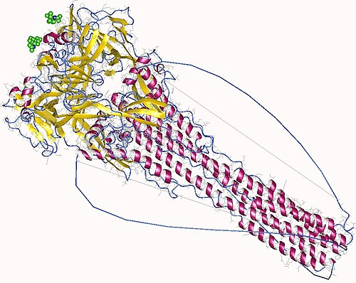
image: By Deposition authors: Yin, H. S., Paterson, R. G., Wen, X., Lamb, R. A., Jardetzky, T. S.;visualization author: User:Astrojan – http://www.rcsb.org/pdb/explore/explore.do?structureId=1ztm, CC BY 4.0, https://commons.wikimedia.org/w/index.php?curid=50142524