Norovirus: Key Facts at a Glance
Norovirus is a highly contagious virus that causes acute gastroenteritis, a sudden inflammation of the stomach and intestines. It is often called the winter vomiting bug or viral stomach flu, although it is unrelated to the influenza virus. Symptoms typically begin 12 to 48 hours after exposure, and most healthy people recover within 1 to 3 days.
- Main symptoms: Severe nausea, sudden projectile vomiting, watery non-bloody diarrhea, and cramping abdominal pain. Low-grade fever, fatigue, muscle aches, chills, and headache can also occur.
- How it spreads: Through direct contact with an infected person, contaminated food or water (such as raw shellfish, unwashed leafy greens, and berries), and contaminated surfaces. Droplets from projectile vomiting can also carry the virus.
- How long it is contagious: People are most contagious while they have vomiting and diarrhea, but the virus continues to shed in stool for 2 to 3 weeks or more after symptoms have cleared. Stay home during the illness and for at least 48 hours after symptoms resolve.
- Treatment: There is no specific antiviral medicine, and antibiotics do not work against it. Care focuses on preventing dehydration with small, frequent sips of oral rehydration solution. Breastfeeding and infant formula should continue without dilution.
- Warning signs in children: Severe lethargy, sunken eyes, a sunken soft spot (fontanelle) in infants, no tears when crying, and fewer than one wet diaper in 6 to 8 hours require urgent pediatric assessment.
- Prevention: Wash hands with soap and running water for at least 20 seconds. Alcohol-based hand sanitizers do not reliably inactivate the virus. Disinfect contaminated surfaces with a chlorine bleach solution, wash fresh produce carefully, and cook shellfish thoroughly.
- Vaccine: No licensed norovirus vaccine is currently available, so careful infection prevention remains the primary defense.
Norovirus Demystified: The Comprehensive Clinical and Epidemiological Guide
Norovirus represents the leading etiology of nonbacterial acute gastroenteritis and foodborne diarrheal disease across the globe. Widely recognized in public health records and popular terminology as the winter vomiting bug, this non-enveloped viral pathogen demonstrates an extraordinary level of infectivity, high environmental stability, and a remarkable capacity to cause explosive outbreaks across both community institutions and health care environments. Understanding how norovirus operates at a molecular, clinical, and immunological level is paramount for medical practitioners, epidemiologists, and caregivers striving to curtail transmission chains and reduce global hospitalization rates.
The medical and socio-economic burden imposed by acute gastrointestinal illness linked to norovirus remains substantial in every corner of the world. Each year, the pathogen triggers tens of millions of severe illness episodes, causing significant numbers of outpatient consultations, emergency admissions, and fatal complications among infants and fragile older people populations. Because an incredibly low infectious dose—ranging from as few as 10 to 100 virions—suffices to trigger productive gastroenteritis, containment demands aggressive infection control, deep surface disinfection, and strict personal hygiene routines.
The Virology, Genomic Architecture, and Diversity of Norovirus
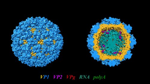
The genus Norovirus belongs to the family Caliciviridae, a distinct family of positive-sense, single-stranded RNA viruses. Originally identified through immunoelectron microscopy following a 1968 outbreak in Norwalk, Ohio, the prototype virion exhibits a distinct morphology measuring roughly 27 to 38 nanometers in diameter. Structurally, the virion lacks a lipid bilayer envelope and is arranged in a classic icosahedral architecture characterized by cup-shaped surface indentations that give the family its name.
Molecular Organization of the Viral Genome
The linear, positive-sense viral RNA genome spans approximately 7.5 to 7.7 kilobases. It terminates in a polyadenylated tail at its 3-prime end and possesses a covalently bound viral protein genome (VPg) cap at its 5-prime terminus, which is indispensable for initiating viral translation within host enterocytes. The coding potential of human norovirus is partitioned into three distinct open reading frames:
- Open Reading Frame 1 (ORF1): Encodes an extensive polyprotein that is proteolytically cleaved by the viral 3C-like protease into six essential nonstructural functional proteins, including the RNA-dependent RNA polymerase, nucleoside triphosphatase (NTPase), p48 (N-terminal protein), and VPg.
- Open Reading Frame 2 (ORF2): Encodes the major structural capsid protein, VP1, which weighs roughly 57 to 60 kilodaltons. Ninety VP1 dimers assemble spontaneously into the icosahedral capsid shell, forming the protective barrier that encloses genomic RNA.
- Open Reading Frame 3 (ORF3): Encodes VP2, a minor basic structural protein that interacts with the inner face of the shell to provide structural stability and support packaging functions.
| Genomic Domain | Encoded Product | Biological and Structural Role |
| ORF1 Polyprotein | Nonstructural Protease, Polymerase, NTPase, VPg | Drives genomic replication, transcription, and translation inside host enterocytes. |
| ORF2 Capsid Shell (S Domain) | Inner structural shell of VP1 | Forms the conserved interior framework of the icosahedral virion. |
| ORF2 Protruding (P Domain: P1 & P2) | Outer protruding arch of VP1 | Mediates attachment to host cellular receptors and bears key neutralization epitopes. |
| ORF3 Minor Protein | Structural VP2 protein | Interacts with VP1 dimers to ensure proper virion packaging and conformational integrity. |
The structural VP1 monomer splits functionally into two distinct domains: the Shell (S) domain and the Protruding (P) domain. The P domain extends upward from the shell and is subdivided into the P1 stalk and the hypervariable P2 domain. Because the P2 domain sits directly on the outermost exposed surface of the virion, it contains the primary cellular receptor-binding interfaces and the immunodominant epitopes recognized by human neutralizing antibodies.
Classification and Evolutionary Lineages of Norovirus
The genus encompasses a broad array of genogroups and genotypes based on nucleotide and amino acid sequence divergence in the VP1 region. To date, noroviruses are classified into at least ten distinct genogroups (designated GI through GX), among which GI, GII, and GIV represent the primary lineages responsible for clinical illness in humans:
- Genogroup I (GI): Contains at least nine genotypes, including the historical prototype Norwalk strain (GI.1).
- Genogroup II (GII): The most prevalent and genetically diverse lineage, encompassing more than twenty-five distinct genotypes.
- Genogroup IV (GIV): Less frequently isolated, known to cause sporadic human enteritis as well as cross-species infections.
Within this expansive phylogeny, norovirus strains belonging to the GII.4 genotype have served as the predominant global pandemic drivers over the past several decades. The GII.4 lineage undergoes rapid epochal evolution, accumulating nonsynonymous mutations in the hypervariable P2 domain. This process mirrors antigenic drift in influenza viruses, enabling novel GII.4 variants to escape herd immunity and sweep through susceptible human populations every two to four years. Major historical pandemic variants include Grimsby (1996), Farmington Hills (2002), Hunter (2004), Den Haag (2006b), New Orleans (2009), and Sydney (2012). In surveillance data spanning norovirus 2025 and ongoing surveillance into norovirus 2026, emergent recombinant strains—including GII.17 Kawasaki, novel GII.2 variants, and GII.4 Sydney recombinants—continue to alter the global epidemiological landscape.
Transmission Dynamics and Epidemiology of Norovirus
The transmission potential of norovirus is unparalleled among gastrointestinal viruses. Due to its resistance to physiological pH changes, lipid solvents, low freezing temperatures, and heat up to 60 degrees Celsius, virions survive for prolonged periods on dry inert objects and within aquatic environments.
| Transmission Step | Vehicle / Mechanism | Resulting Exposure in Host |
| Fecal-Oral Vector | Unwashed hands, poor diaper hygiene, contaminated utensils | Ingestion of virions through direct hand-to-mouth contact. |
| Foodborne Vehicle | Raw shellfish (oysters), unwashed leafy greens, berries | Virions concentrated in gastrointestinal tract during digestion. |
| Waterborne Vehicle | Contaminated drinking wells, public pools, municipal ice supplies | Direct ingestion of high-titer sewage-impacted water. |
| Fomite Surface | Doorknobs, handrails, electronic touchscreens, linens | Transfer of resilient virions from non-porous surfaces to mouth. |
| Aerosolized Droplets | High-velocity projectile vomiting in indoor spaces | Direct inhalation and involuntary swallowing of viral droplets. |
Primary Modes of Transmission
Human-to-human transmission operates through multiple intertwined routes:
- The Direct Fecal-Oral Route: Unwashed or improperly sanitized hands serve as direct vectors for viral transport from fecal residue to the mouth or food preparation surfaces.
- Foodborne Transmission: Consuming food contaminated during agricultural harvesting, packaging, or kitchen preparation represents a major cause of extensive community outbreaks. Filter-feeding shellfish, particularly oysters, clams, and mussels cultivated in sewage-impacted coastal waters, concentrate viral particles within their digestive diverticula. Raw produce, such as unwashed leafy greens, berries, and salads, is frequently implicated.
- Waterborne Transmission: Municipal water system breakdowns, private wells, recreational swimming pools, and commercial ice supplies contaminated with sewage can expose broad cohorts to high viral loads.
- Fomite and Environmental Surfaces: Fecal matter or expelled vomitus coats environmental fomites such as doorknobs, handrails, mobile electronics, bathroom fixtures, and linens. Inadvertent contact followed by hand-to-mouth behaviors triggers productive infection.
- Aerosolized Droplets from Vomitus: Projectile emesis shears gastrointestinal fluids into microscopic droplets that travel through indoor air. Nearby individuals can inhale and swallow these droplets or touch surfaces where they settle.
High-Risk Transmission Environments
Outbreaks flourish where individuals gather in dense, enclosed environments with shared sanitation facilities and dining areas:
- Health Care Facilities and Nursing Homes: Long-term care institutions and hospital wards report the highest frequency of prolonged norovirus outbreaks. Vulnerable older people residents face heightened clinical risks.
- Cruise Ships and Hospitality Settings: Closed vessel architecture and shared buffets facilitate rapid transmission across thousands of passengers within days.
- Schools, Child Care Centers, and Universities: High contact rates and variable personal hand hygiene among young children create ideal conditions for explosive outbreaks.
- Military Garrisons and Operational Fields: Crowded field conditions and close living quarters frequently result in high attack rates among active-duty personnel.
Genetic Susceptibility and the Host Receptor Paradigm
Host susceptibility to norovirus is not uniform across human populations. Breakthroughs in molecular biology and host glycobiology have revealed that attachment and viral entry into human enterocytes depend heavily on interactions with histo-blood group antigens (HBGAs).
The Role of FUT2, FUT3, and ABO Loci
HBGAs are complex oligosaccharide motifs adorning glycoproteins and glycolipids on mucosal surfaces and within body fluids such as saliva and digestive secretions. The enzymatic biosynthesis of these structures involves sequential monosaccharide additions controlled by specific genes:
Biosynthetic Pathway of Mucosal Receptors
- Foundational Precursor: Precursor oligosaccharide chains form on the apical enterocyte surface.
- FUT2 Gene Action (Secretor Status): The alpha-1,2-fucosyltransferase enzyme adds fucose to synthesize the essential H type 1 antigen.
- ABO Glycosyltransferase Action: Depending on host genotype, specific enzymes add N-acetylgalactosamine (Blood Group A) or galactose (Blood Group B) to the H antigen structure.
- FUT3 Gene Action (Lewis Status): Alpha-1,4-fucosyltransferase synthesizes Lewis a and Lewis b oligosaccharide chains.
Individuals who possess at least one functional allele of the FUT2 gene (encoding alpha-1,2-fucosyltransferase) synthesize the foundational H type 1 antigen on mucosal surfaces and secrete it into bodily fluids. These individuals are designated secretor-positive. The ABO glycosyltransferases then attach an N-acetylgalactosamine (yielding the A antigen) or a galactose (yielding the B antigen) onto this fucosylated H core. Meanwhile, the FUT3 gene encodes an alpha-1,4-fucosyltransferase that synthesizes the Lewis a and Lewis b oligosaccharide structures.
Secretor Status and Natural Resistance
Approximately twenty percent of individuals of European and African descent carry homozygous nonsense mutations in the FUT2 coding sequence—most notably the 428G-to-A nonsense mutation—that eliminate fucosyltransferase activity. These individuals are classified as nonsecretors (FUT2 -/-).
Clinical Research Insight
Human challenge studies and epidemiological field analyses have demonstrated that homozygous nonsecretors are naturally resistant to infection and illness caused by Norwalk virus (GI.1) and predominant pandemic GII.4 variants, because their intestinal enterocytes lack the alpha-1,2-fucosylated docking structures required for viral capsid attachment.
| Phenotypic Status | Genetic Profile | Mucosal HBGA Expression | Susceptibility Profile to Norovirus Strains |
| Homozygous Secretor | FUT2 +/+ | Abundant H type 1, Lewis b, A/B antigens | Highly susceptible to GII.4, GI.1, and common pandemic strains. |
| Heterozygous Secretor | FUT2 +/- | Expresses alpha-1,2-linked fucose antigens | Susceptible to all common secretor-dependent lineages. |
| Homozygous Nonsecretor | FUT2 -/- (e.g., 428G>A) | Lacks alpha-1,2 fucose; displays Lewis a | Resistant to GII.4 and GI.1; vulnerable to secretor-independent strains (GI.3, GII.2). |
| Weak-Secretor | Se385 Missense Variant | Attenuated mucosal H antigen density | Common in East Asia; shows partial protection against GII.4 with moderate susceptibility. |
This Mendelian resistance model applies primarily to secretor-dependent lineages. Certain other genotypes, including GI.3, GII.2 (Snow Mountain virus), and GII.7, infect secretors and nonsecretors alike. These genotypes utilize alternative glycan motifs or secondary protein receptors that do not depend strictly on alpha-1,2-linked mucosal fucose.
Pathophysiology and Mechanisms of Mucosal Injury
Once swallowed, the norovirus virion traverses the acidic stomach lumen, protected by its non-enveloped capsid shell. Upon entering the proximal small bowel—predominantly the jejunum and upper duodenum—the capsid P2 domain attaches to mucosal HBGAs.
Cellular Tropism and Enteric Replication
For decades, the inability to cultivate human noroviruses in classic cell culture obscured the precise identity of host target cells. Breakthroughs using stem-cell-derived human intestinal enteroids (HIEs) and human-induced pluripotent stem cell-derived intestinal epithelial systems demonstrated that human norovirus readily infects mature enterocytes lining the intestinal villi. Commensal gut bacteria expressing HBGA-like antigens can also bind noroviral capsids, facilitating viral presentation and infection of underlying gut-associated lymphoid cells.
| Pathophysiological Phase | Cellular Event | Clinical Impact |
| Phase 1: Attachment & Entry | Capsid P2 domain binds apical HBGAs on villus enterocytes. | Virion endocytosis and initiation of viral disassembly. |
| Phase 2: Viral Replication | Translation of ORF1 proteins; RNA replication. | Enterocyte shutdown and cellular cytotoxic damage. |
| Phase 3: Epithelial Remodeling | Apoptosis causes villus blunting and crypt hyperplasia. | Marked reduction in total absorptive surface area. |
| Phase 4: Functional Breakdown | Brush-border enzyme depletion; tight junction leakage. | Osmotic and secretor-driven watery diarrhea. |
| Phase 5: Neurohormonal Trigger | Enteric inflammation triggers serotonin (5-HT) release. | Stimulation of medullary emetic centers causing sudden emesis. |
Mechanisms of Diarrhea and Emesis
The cellular replication cycle induces targeted histopathological abnormalities while leaving the deep intestinal mucosa intact:
- Villus Blunting and Broadening: The infection triggers apoptosis, enterocyte rounding, and cytoplasmic vacuolation, leading to marked shortening of the absorptive villi in the jejunum.
- Enzymatic Depletion: Expression of apical brush-border digestive enzymes—including alkaline phosphatase, sucrase, and trehalase—drops substantially, impairing carbohydrate absorption and triggering osmotic fluid shifts into the intestinal lumen.
- Increased Tight Junction Permeability: Epithelial barrier disruption and zonula occludens relocation allow passive leakage of electrolytes and systemic water into the gut lumen, causing watery diarrhea.
- Delayed Gastric Emptying: Intestinal inflammation and neurohormonal serotonin (5-HT) release stimulate the area postrema and emetic centers in the medulla, inducing sudden projectile emesis.
Clinical Picture and Symptoms
The presentation of acute norovirus gastroenteritis is characterized by an abrupt onset following a brief incubation period. Patients often shift from complete wellness to severe gastrointestinal distress within hours.
Incubation Period and Symptom Progression
The incubation window typically spans 12 to 48 hours following viral ingestion, with a median of 24 to 36 hours. The hallmark clinical signs include:
- Severe, Recurrent Nausea: Often serves as the earliest warning sign.
- Sudden, Projectile Vomiting: Particularly prominent in pediatric patients and young adults.
- Watery Diarrhea: Copious, non-bloody, loose liquid stools without inflammatory leukocytes or macroscopic mucosal tissue.
- Cramping Abdominal Pain: Diffuse, intermittent, spasmodic periumbilical discomfort.
- Systemic Manifestations: Low-grade fever, general fatigue, myalgias, shivering chills, and severe throbbing headache.
For the majority of previously healthy individuals, acute symptoms peak rapidly and resolve within 24 to 72 hours without permanent gastrointestinal injury.
Pediatric and Geriatric Complications
Although norovirus is self-limiting in healthy adults, it causes severe complications at the extremes of age:
Clinical Warning: High-Risk Populations
- Pediatric Red Flags: Severe lethargy, sunken eyes, depressed fontanelles in infants, absent tears when crying, and anuria (fewer than one wet diaper in 6 to 8 hours) require urgent pediatric assessment.
- Geriatric Complications: Rapid volume contraction triggers orthostatic dizziness, prerenal azotemia, cognitive disorientation, fall-related trauma, and decompensation of underlying cardiovascular disease.
In neonates and preterm infants, the infection can trigger intestinal ischemia and has been linked to necrotizing enterocolitis in neonatal intensive care units. Among adults aged 65 and older, severe volume loss can precipitate electrolyte derangements, acute tubular necrosis, cardiovascular collapse, and elevated all-cause mortality.
Persistent Infection in the Immunocompromised
Patients with primary or acquired immunodeficiencies face substantially altered disease courses:
- Solid Organ Transplant Recipients: Patients on calcineurin inhibitors or antimetabolite immunosuppressants can develop chronic norovirus enteritis that persists for months to years, causing malnutrition and allograft dysfunction.
- Hematopoietic Stem Cell Transplant Recipients: Chronic diarrheal shedding can complicate post-transplant care, occasionally mimicking or compounding gastrointestinal graft-versus-host disease.
- Long-Term Shedding: While immunocompetent individuals clear the virus within 2 to 3 weeks, immunocompromised patients can excrete replication-competent virions in their stool for years, acting as long-term environmental reservoirs.
Diagnostic Methodologies and Laboratory Identification
Because symptoms of acute gastroenteritis can arise from multiple viral, bacterial, or parasitic agents, clinical evaluation alone cannot definitively identify norovirus without diagnostic testing.
| Diagnostic Tool | Specimen Type | Typical Turnaround Time | Clinical Sensitivity & Specificity | Best Practical Setting |
| Real-Time RT-PCR | Stool (or vomitus) | 2 to 4 hours | Gold standard: Sensitivity >95%, Specificity >99% | Definitive clinical diagnosis and outbreak strain genotyping. |
| Syndromic Multiplex PCR | Diarrheal stool | 1 to 2 hours | High: Sensitivity >92%, Specificity >98% | Rapid emergency and hospital screening across multiple pathogens. |
| Enzyme Immunoassay (EIA) | Bulk stool specimen | 1 to 2 hours | Moderate: Sensitivity 30-80%, Specificity 85-99% | Outbreak screening where at least 6 samples are collected. |
| Rapid Lateral Flow Strips | Fresh stool suspension | 15 to 30 minutes | Variable: Sensitivity 20-80%, Specificity >90% | Point-of-care triaging; negative results require confirmation. |
Kaplan’s Clinical and Epidemiologic Criteria
Prior to the availability of molecular testing, epidemiologists relied on Kaplan’s criteria to identify suspected norovirus outbreaks during investigations:
- Vomiting is reported by more than 50% of affected patients.
- The mean or median incubation period ranges from 24 to 48 hours.
- The mean or median duration of illness spans 12 to 60 hours.
- Routine stool cultures are negative for bacterial enteropathogens.
While Kaplan’s criteria remain helpful for initial field assessments, modern clinical decision-making relies on molecular assays.
Molecular Testing: The Diagnostic Gold Standard
Reverse transcriptase-polymerase chain reaction (RT-PCR) serves as the benchmark for detecting and typing norovirus in clinical and public health laboratories:
- Target Regions: Real-time RT-PCR assays primarily target the conserved junction between ORF1 and ORF2. This boundary region is well conserved across diverse genogroups, providing high clinical sensitivity for both GI and GII strains.
- Multiplex Syndromic GI Panels: Comprehensive panels allow simultaneous detection of noroviruses alongside rotaviruses, astroviruses, adenoviruses, and bacterial pathogens within 1 to 2 hours directly from stool specimens.
- Isothermal Amplification: Methods such as RT-loop-mediated isothermal amplification (RT-LAMP) and nucleic acid sequence-based amplification (NASBA) offer rapid results with simpler instrumentation.
Antigen Detection: Immunoassays
Commercial enzyme immunoassays (EIAs) and rapid lateral flow immunochromatographic strips detect VP1 capsid antigens directly in stool specimens. While these kits provide results within 15 to 30 minutes, their sensitivity ranges from 30% to 80% compared to real-time RT-PCR, due to sequence divergence across strains. Public health guidelines therefore recommend testing at least six independent stool samples from an outbreak before ruling out norovirus based on negative antigen assays.
Medical Treatment, Supportive Care, and Novel Therapeutics
There are no approved, pathogen-specific antiviral therapies that cure norovirus. Management remains centered on supportive care, rapid rehydration, and preventing fluid and electrolyte depletion.
Clinical Pathway: Triage and Rehydration
- Mild to Moderate Illness: Administer commercial Oral Rehydration Salts (ORS) in small, frequent sips to leverage sodium-glucose enterocyte cotransporters.
- Severe Illness: Initiate rapid intravenous crystalloid infusion (normal saline or lactated Ringer’s) when emesis is intractable, dehydration is severe, or hemodynamic instability is present.
Oral and Intravenous Rehydration Strategies
Fluid replacement must begin as soon as symptoms develop:
- Oral Rehydration Therapy (ORS): Commercial ORS formulations containing precise ratios of glucose and sodium leverage sodium-glucose cotransporters in the small intestine, enhancing water absorption across undamaged enterocytes. Fluids should be consumed in small, frequent sips to avoid triggering emesis.
- Beverages to Avoid: Clear sodas, apple juice, undiluted sports beverages, and hypertonic drinks contain excessive simple sugars that can worsen osmotic diarrhea. Caffeinated and alcoholic beverages should be avoided due to their diuretic properties.
- Intravenous (IV) Resuscitation: When continuous emesis prevents oral fluid retention, or when signs of severe dehydration emerge—such as profound hypotension, oliguria, tachycardia, or altered mental status—intravenous crystalloid infusion (normal saline or lactated Ringer’s) is required to restore intravascular volume.
Dietary Modifications and Nutrition
Patients should transition back to solid foods as tolerated once vomiting subsides:
- The BRAT Diet: Bananas, rice, applesauce, and toast were once recommended during recovery, but the American Academy of Pediatrics (AAP) no longer considers this diet useful. Because these foods are low in fiber, protein, and fat, the diet does not supply enough nutrition to help a child’s digestive tract recover, and some pediatricians believe it may make symptoms last longer. Once clear liquids stay down, the AAP advises offering a few small bites at a time of easy-to-digest foods without seasoning, such as fruits, vegetables, low-sugar yogurt, or whole-grain bread. Children can then return to a normal, well-balanced diet appropriate for their age, including fruits, vegetables, meat, yogurt, and complex carbohydrates.
- Foods to Limit: Dairy products containing lactose, greasy preparations, and spicy meals should be temporarily withheld until brush-border digestive enzymes recover.
- Pediatric Feeding: Breastfeeding and infant formula regimens should continue without dilution. Formula should be offered in smaller, more frequent volumes to maintain caloric intake.
Pharmacological Considerations and Experimental Agents
Medications should be used selectively under clinical supervision:
- Antiemetic Agents: Ondansetron, a selective 5-HT3 receptor antagonist, may be prescribed in hospital or emergency settings to control intractable emesis, making oral rehydration feasible.
- Antimotility Agents: Over-the-counter antimotility medications such as loperamide may reduce stool frequency in non-febrile adults, but they are contraindicated in young children and in cases of invasive, inflammatory, or bloody diarrhea.
- Antibiotic Use: Because norovirus is viral, antibacterial agents are entirely ineffective. Inappropriate antibiotic administration disrupts commensal gut microbiota and can exacerbate diarrhea.
- Investigational Antivirals: In chronic, refractory cases in immunocompromised patients, nitazoxanide and enteral human immunoglobulin have shown modest benefits in case reports, but broad efficacy has not been demonstrated in clinical trials.
Infection Control and Prevention Protocols
Because norovirus sheds in extraordinarily high concentrations and resists standard alcohol-based cleaners, breaking transmission chains requires strict adherence to infection control guidelines.
| Prevention Domain | Recommended Protocol | Critical Clinical / Practical Detail |
| Hand Hygiene | Running water and soap lathering for at least 20 seconds. | Hand rubs containing alcohol are insufficient for non-enveloped virions. |
| Surface Sanitization | Sodium hypochlorite (chlorine bleach) solution. | 1,000 ppm for surfaces; 5,000 ppm for direct vomit or stool spills. |
| Food Safety | Thorough cooking of shellfish; washing raw produce. | Shellfish steaming must reach internal temperatures above 90°C. |
| Linens & Laundry | Separate handling and washing at high temperatures. | Wash soiled bedding and clothes at a minimum temperature of 60°C. |
| Exclusion Rules | Strict workplace and school absence policies. | Stay home during illness and for at least 48 hours after symptoms resolve. |
The Inadequacy of Alcohol-Based Sanitizers
A key factor in institutional norovirus outbreaks is over-reliance on alcohol-based hand rubs. Because the non-enveloped capsid lacks a vulnerable lipid bilayer, ethanol- and isopropanol-based formulations alone do not reliably inactivate the virus.
Essential Infection Control Standard
Vigorous mechanical handwashing using running water and soap for at least 20 seconds remains the gold standard for removing norovirus virions from the skin surface. Hand friction physically dislodges viral particles, which are then rinsed away down the drain.
Surface Disinfection with Chlorine Bleach
Standard household cleansers and quaternary ammonium compounds do not reliably neutralize norovirus on non-porous surfaces. Instead, environmental surfaces must be disinfected using chlorine bleach (sodium hypochlorite) solutions:
- Routine Surface Sanitation: A chlorine concentration of 1,000 parts per million (roughly a 1:50 dilution of standard 5.25% household bleach) is recommended for high-touch, non-porous surfaces.
- Vomitus and Fecal Decontamination: For direct spills of emesis or stool, a higher concentration of 5,000 parts per million (a 1:10 dilution of standard household bleach) is required.
- Application Technique: Disinfection teams should wear disposable gloves, blot gross fluids with absorbent disposable paper towels, apply the bleach solution generously, allow a minimum contact time of 5 to 10 minutes, and then rinse or allow to air dry.
- Laundry Handling: Soiled clothing and bed linens should be transported in closed bags, handled with minimal agitation to prevent aerosolization, and washed in hot cycles (at least 60 degrees Celsius) using appropriate bleach-safe detergents.
Exclusion Policies in Institutions
Infected food service employees, nurses, clinicians, and day care workers should be strictly excluded from their workstations during their symptomatic illness and for at least 48 hours after vomiting and diarrhea have completely resolved. In hospitals, affected wards often require cohort isolation, visitor restrictions, and admission freezes to prevent transmission to adjacent units.
The Landscape of Norovirus Vaccines
Developing a licensed norovirus vaccine remains an active area of public health research. The economic and societal rationale is clear: a moderately effective prophylactic vaccine could prevent millions of medical visits and save billions of dollars in health care expenses annually.
Barriers to Vaccine Development
Several biological features of norovirus complicate vaccine development:
- Broad Antigenic Diversity: With multiple genogroups, dozens of genotypes, and ongoing evolution within GII.4, vaccines must generate cross-protective immunity across divergent strains.
- Short-Lived Natural Immunity: Protective immunity following natural infection can be relatively transient, lasting from several months to a few years, after which individuals may become susceptible to homologous or heterologous reinfection.
- Host Glycan Polymorphism: Differences in mucosal HBGA presentation across human populations mean that vaccine responses must be effective across diverse genetic backgrounds, including both secretors and weak-secretors.
- Historical Inability to Propagate the Virus: Decades without an in vitro neutralization assay made it difficult to assess protective antibodies in preclinical models.
Virus-Like Particle (VLP) Platforms
Most vaccine candidates in clinical development utilize recombinant virus-like particles (VLPs). When the VP1 structural capsid gene is expressed in insect or mammalian cell cultures, it spontaneously self-assembles into empty capsids that mimic the natural virion’s size and antigen presentation, without containing viral RNA.
- Intranasal and Intramuscular Formulations: Clinical trials evaluating bivalent VLP candidates containing GI.1 and GII.4 antigens combined with adjuvants (such as monophosphoryl lipid A and aluminum hydroxide) have elicited elevated serum blocking antibodies and reduced the incidence and severity of illness following experimental challenges.
- Future Outlook: Candidate vaccines continue to progress through phase 2 and phase 3 clinical evaluations. Until a broadly effective formulation is approved and distributed globally, meticulous infection prevention remains the primary defense against norovirus transmission.
Comparison: Norovirus vs. Rotavirus
While both norovirus and rotavirus cause acute viral gastroenteritis, distinct differences in their virology, clinical presentation, and epidemiology differentiate the two conditions.
| Clinical & Epidemiological Characteristic | Norovirus Infection | Rotavirus Infection |
| Viral Family & Genome | Caliciviridae; positive-sense, single-stranded RNA. | Reoviridae; segmented, double-stranded RNA. |
| Primary Target Age Group | Individuals of all ages; high impact in adults and the older people. | Predominantly infants and young children under 5 years. |
| Dominant Clinical Symptom | Explosive emesis and sudden vomiting; watery diarrhea. | Severe, dehydrating watery diarrhea; vomiting less dominant. |
| Protective Prophylactic Vaccine | No licensed vaccine currently available worldwide. | Live oral vaccines widely integrated into immunization schedules. |
| Environmental Stability | Exceptionally resistant to freezing, heat (60°C), and alcohol. | Moderately stable, but more susceptible to standard hospital disinfectants. |
| Incubation Window | Rapid: typically 12 to 48 hours. | Longer: typically 48 to 72 hours. |
| Post-Illness Shedding | Can continue in stool for 2 to 4 weeks or longer. | Typically clears within 7 to 10 days post-recovery. |
Frequently Asked Questions
What is norovirus?
Norovirus is a highly contagious, non-enveloped RNA virus from the Caliciviridae family that causes acute gastroenteritis, marked by sudden inflammation of the stomach and intestines. It is often referred to in public health contexts as the winter vomiting bug or viral stomach flu, although it is unrelated to the influenza virus. The virus affects individuals across all age brackets and represents the leading cause of nonbacterial gastrointestinal outbreaks globally.
How long does norovirus last?
For most healthy individuals, acute symptoms of norovirus infection typically resolve within 1 to 3 days (24 to 72 hours). However, the illness may persist considerably longer—often lasting up to a week—in neonates, young children, and older adults. In severely immunocompromised individuals, such as transplant recipients receiving immunosuppressive medications, the infection can become chronic, causing persistent symptoms and shedding that last for months or even years.
How long is norovirus contagious?
Individuals infected with norovirus are most contagious during the acute symptomatic phase when they are actively experiencing vomiting and diarrhea. However, the virus continues to shed in feces in high concentrations for 2 to 3 weeks or more after all symptoms have cleared. Because shedding persists well into the recovery period, practicing meticulous hand hygiene and observing institutional exclusion policies for at least 48 hours after symptoms resolve is essential to interrupt transmission.
How is norovirus spread?
Norovirus spreads primarily via the fecal-oral route through direct contact with an infected person, such as caring for a sick family member or sharing utensils. It is also transmitted by consuming food or water contaminated at the source or during preparation, and by touching contaminated surfaces before touching the mouth. Furthermore, because projectile vomiting can aerosolize virus particles into microscopic droplets, the virus can spread when nearby individuals inhale or swallow these droplets or touch surfaces where they settle.
How to treat norovirus?
There are no specific antiviral medications or antibiotic therapies that eliminate a norovirus infection. Management relies on supportive care, with the primary objective being the prevention and correction of dehydration and electrolyte imbalances. Mild to moderate dehydration is managed with small, frequent sips of oral rehydration solutions, while severe cases marked by intractable emesis or hypovolemic shock require hospitalization for intravenous fluid resuscitation.
What is norovirus prevention?
Preventing norovirus relies on rigorous personal hand hygiene, thorough environmental decontamination, and safe food handling practices. Hands should be washed thoroughly with soap and warm running water for at least 20 seconds, as alcohol-based hand sanitizers do not reliably inactivate this non-enveloped virus. Surfaces potentially contaminated with vomit or feces should be disinfected using a chlorine bleach solution (1,000 to 5,000 ppm), while fresh produce should be washed carefully, bivalve shellfish thoroughly cooked, and sick individuals excluded from food preparation for at least 48 hours after recovery.
The following posts may interest you
Adenovirus: A Complete Guide for Parents and Kids
Hantavirus Symptoms: Understanding the Major Risks
Rhinovirus Infections in Children: Causes, Risks, and Care
Rotavirus: Symptoms, Causes, Vaccine and Care Guide
Disclaimer: The content provided on BabyKidsHealth.com is for informational purposes only and is not intended as a substitute for professional medical advice, diagnosis, or treatment. Always seek the advice of your physician or other qualified health provider with any questions you may have regarding a medical condition.
Sources
About Norovirus (Centers for Disease Control and Prevention, CDC)
https://www.cdc.gov/norovirus/about/index.html
How Norovirus Spreads (Centers for Disease Control and Prevention, CDC)
https://www.cdc.gov/norovirus/causes/index.html
How to Prevent Norovirus (Centers for Disease Control and Prevention, CDC)
https://www.cdc.gov/norovirus/prevention/index.html
Norovirus: What to Do If Your Child Catches This Contagious “Stomach Bug” (American Academy of Pediatrics, HealthyChildren.org)
Diarrhea in Children: What Parents Need to Know (American Academy of Pediatrics, HealthyChildren.org)
https://www.healthychildren.org/English/health-issues/conditions/abdominal/Pages/Diarrhea.aspx
Norovirus (World Health Organization, WHO)
https://www.who.int/teams/immunization-vaccines-and-biologicals/diseases/norovirus
Norovirus
https://journals.asm.org/doi/full/10.1128/cmr.00075-14
Mendelian resistance to human norovirus infections
https://www.sciencedirect.com/science/article/pii/S1044532306001035
image: By Dr. Victor Padilla-Sanchez, PhD – Own work, CC BY-SA 4.0, https://commons.wikimedia.org/w/index.php?curid=193527903