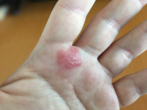
Contact Dermatitis: Key Facts at a Glance
Contact dermatitis is an inflammatory skin rash caused by direct contact with a substance that either damages the skin’s protective outer barrier or triggers a delayed allergic reaction. It often affects the hands and face, and it is not contagious: it cannot spread from person to person, and the fluid inside the blisters does not spread the rash.
- Two main types: Irritant contact dermatitis, caused by direct chemical or physical damage to the skin, accounts for roughly eighty percent of cases and can happen to anyone. Allergic contact dermatitis, about twenty percent of cases, is a delayed immune reaction that develops only after the skin has been sensitized to a specific substance.
- Main symptoms: Allergic reactions mainly cause intense itching, while irritant reactions more often burn, sting, or feel sore. The rash may be red and swollen, with small bumps or blisters that ooze and crust. Allergic rashes usually appear 24 to 48 hours after contact and can spread past the contact area. With ongoing exposure, the skin becomes dry, thickened, and cracked.
- Common triggers: Soaps, detergents, solvents, and frequent wet work; nickel in jewelry, belt buckles, and denim buttons; fragrances and preservatives in cosmetics, shampoos, and wet wipes; hair dyes and black henna tattoos (PPD); and poison ivy, poison oak, and poison sumac.
- Diagnosis: A doctor examines the pattern of the rash and asks about products, hobbies, work, and recent exposures. Patch testing on the upper back, with readings at 48 hours and again at 72 to 96 hours, shows which substance causes an allergic reaction.
- Treatment: Identifying and avoiding the trigger is the foundation of care. Cool compresses and prescribed corticosteroid ointments or creams calm the rash, with milder products or non-steroid creams (tacrolimus, pimecrolimus) for the face and eyelids. Severe or widespread reactions may need oral steroids that a doctor tapers gradually.
- When to see a doctor: Seek medical care if the rash is severe or widespread, causes marked facial swelling, or does not improve after the trigger is avoided and treatment is used. Pus or thick, honey-colored crusts can mean a bacterial skin infection.
- Prevention: Rinse the skin with lukewarm water and a gentle cleanser within 10 to 15 minutes of contact with an irritant or plant oil, wash contaminated clothing and pets, choose fragrance-free cleansers, and apply a thick moisturizer right after washing while the skin is still slightly damp.
Clinical Masterclass on Contact Dermatitis: Pathophysiology, Diagnosis, and Management
Cutaneous inflammation triggered by direct environmental exposure represents one of the most prevalent clinical challenges in contemporary dermatology and occupational health. Among these widespread inflammatory conditions, Contact Dermatitis stands out as an acute or chronic condition caused by skin contact with specific external physical, chemical, or biological agents. Epidemiological analyses demonstrate that inflammatory skin diseases account for a substantial proportion of all work-related illnesses in developed nations, with eczematous skin eruptions responsible for significant morbidity, time lost from employment, and profound psychosocial distress. Clinically, Contact Dermatitis manifests along a broad spectrum ranging from mild, localized erythematous macules and subtle desquamation to severe, widespread blistering eruptions and chronic, disfiguring lichenification. Because its clinical features can mimic various endogenous eczemas, achieving a precise diagnosis requires a thorough understanding of the mechanisms of epidermal barrier disruption, innate immune activation, and delayed-type hypersensitivity. An accurate clinical evaluation enables healthcare professionals to implement targeted interventions that restore barrier integrity, relieve uncomfortable symptoms, and prevent long-term recurrence.
Defining the Scope of Contact Dermatitis
The broad clinical diagnosis of Contact Dermatitis encompasses two distinct pathophysiological categories: irritant contact dermatitis and allergic contact dermatitis. While both conditions culminate in an eczematous cutaneous reaction, their underlying cellular mechanisms, immunological pathways, and clinical courses differ markedly. Irritant contact dermatitis is the most frequent presentation, accounting for roughly eighty percent of all diagnosed cases. It occurs as a non-immunologic reaction resulting from direct physical or chemical damage to the protective stratum corneum and underlying epidermal keratinocytes. Because it does not rely on antigen-specific immune priming, irritant contact dermatitis can develop in any individual exposed to an irritant of sufficient concentration and duration.
Conversely, allergic contact dermatitis comprises approximately twenty percent of cases and represents an antigen-specific, delayed-type cell-mediated hypersensitivity reaction. It develops only in individuals who have undergone prior immunologic sensitization to a specific hapten. Subsequent re-exposure activates memory T lymphocytes within the skin, sparking an inflammatory cascade. In real-world clinical practice, overlapping phenotypes are common. Chronic exposure to low-grade irritants often disrupts the stratum corneum, facilitating the penetration of environmental haptens that drive secondary allergic contact dermatitis. Recognizing whether a patient presents with an irritant reaction, an allergic reaction, or a mixed dermatitis contact dermatitis presentation is the cornerstone of effective clinical management.
Clinical Practice Insight: Distinguishing Eczematous Reaction Patterns
- Irritant Contact Dermatitis: A non-immunological reaction caused by direct toxic or chemical injury to the stratum corneum, characterized by immediate or cumulative burning, sharp borders, and absence of prior sensitization.
- Allergic Contact Dermatitis: An immunologically driven type IV hypersensitivity response mediated by hapten-specific T cells, characterized by intense pruritus, a delayed crescendo onset (24 to 72 hours post-contact), and spreading borders.
Epidermal Barrier Mechanics in Contact Dermatitis
The stratum corneum is the human body’s primary defense against external environmental hazards. It is organized in a classic brick-and-mortar architecture, where protein-rich, non-nucleated corneocytes are embedded within a hydrophobic lipid matrix composed of ceramides, free fatty acids, and cholesterol. This specialized lipid barrier limits transepidermal water loss and blocks the penetration of external xenobiotics and biological pathogens. In irritant Contact Dermatitis, chemical agents such as surfactants, organic solvents, and alkaline substances dissolve and extract these intercellular lipids, damaging corneocyte membranes and denaturing intracellular keratin.
Following this physical disruption, viable epidermal keratinocytes act as active signal transducers. Physical cellular stress triggers the immediate release of pre-formed alarmins and primary inflammatory cytokines, notably interleukin-1 alpha (IL-1α), interleukin-1 beta (IL-1β), and tumor necrosis factor-alpha (TNF-α). These primary cytokines stimulate adjacent vascular endothelial cells and resident dermal fibroblasts, driving the expression of vascular adhesion molecules and the secretion of chemokines such as CXCL8 and CCL20. This biochemical cascade draws circulating neutrophils and monocytes directly into the affected cutaneous site, producing visible tissue erythema, localized edema, and cellular necrosis. If exposure to the offending chemical is repeated before the barrier can recover, the skin develops cumulative Contact Dermatitis, characterized by chronic inflammation and hyperkeratosis.
Immunopathogenesis of Allergic Contact Dermatitis
Allergic contact hypersensitivity requires a tightly orchestrated immunological process divided into two primary phases: sensitization and elicitation. Contact allergens are typically low-molecular-weight, chemically reactive haptens that penetrate the stratum corneum. Because haptens are too small to provoke an immune response on their own, they must bind covalently to structural carrier proteins in the epidermis to form stable, immunogenic hapten-protein complexes. At the same time, these chemicals stimulate innate immune signaling by generating reactive oxygen species, triggering Toll-like receptors such as TLR4, and activating the intracellular NLRP3 inflammasome within keratinocytes and resident dendritic cells.
Immunological Sequence: From Sensitization to Elicitation
- Phase 1 (Sensitization): Epicutaneous hapten penetration, covalent hapten-carrier protein binding, uptake by epidermal Langerhans cells, migration via afferent lymphatics to regional draining lymph nodes, and antigen presentation to naive T lymphocytes, yielding a pool of circulating memory T cells.
- Phase 2 (Elicitation): Hapten re-exposure on sensitized skin, rapid antigen presentation by local cutaneous dendritic cells, activation of tissue-resident memory T cells (TRM), release of interferon-gamma and interleukin-17, and recruitment of inflammatory leukocytes leading to spongiotic vesiculation.
Specialized antigen-presenting cells, including epidermal Langerhans cells and dermal dendritic cells, internalize these hapten-protein complexes, process them, and upregulate major histocompatibility complex class I and class II molecules along with costimulatory molecules such as CD80 and CD86. These mature dendritic cells migrate through afferent lymphatic channels to regional draining lymph nodes. Within the paracortical areas, they present the processed antigens to naive T lymphocytes. This priming event drives clonal expansion and the differentiation of antigen-specific effector and memory T cells, specifically interferon-gamma-producing CD8+ cytotoxic T cells, CD4+ T helper 1 (Th1) cells, and Th17 cells, which then enter the systemic circulation.
The elicitation phase occurs upon subsequent epicutaneous contact with the same hapten. Cutaneous antigen-presenting cells present the hapten-protein conjugate directly to recirculating memory T cells and tissue-resident memory T (TRM) cells persisting in the dermis and epidermis. Within 24 to 48 hours, these activated memory lymphocytes release large quantities of pro-inflammatory cytokines, including interferon-gamma, interleukin-17, and tumor necrosis factor. This influx triggers intercellular edema between adjacent keratinocytes (spongiosis), microvesicle formation, and the clinical symptoms of an allergic contact dermatitis rash.
| Diagnostic Criterion | Irritant Contact Dermatitis | Allergic Contact Dermatitis |
| Primary Mechanism | Non-immunological physical or chemical barrier destruction | Type IV cell-mediated delayed hypersensitivity reaction |
| Sensitization Requirement | None; occurs on initial exposure to potent agents | Mandatory asymptomatic sensitization period (days to years) |
| Onset Kinetics | Rapid or cumulative; decrescendo pattern post-exposure | Delayed (24 to 72 hours); classic crescendo flare pattern |
| Dominant Symptoms | Stinging, burning, tenderness, and localized cutaneous pain | Intense, unremitting pruritus (itching) |
| Border Morphology | Well-demarcated margins confined strictly to contact zones | Indistinct, spreading borders extending past contact areas |
| Cutaneous Lesions | Erythema, dry scaling, chapping, superficial erosions | Spongiotic papules, edematous plaques, tense vesicles, oozing |
| Diagnostic Confirmation | Clinical diagnosis of exclusion; negative epicutaneous patch test | Positive epicutaneous diagnostic patch test with clinical relevance |
Symptoms and Morphological Phases of Contact Dermatitis
The clinical presentation of Contact Dermatitis varies considerably based on the duration of exposure, the anatomical site involved, and whether the reaction is acute, subacute, or chronic. Patients typically seek medical care because of physical discomfort, itching, and cosmetic concerns. In allergic presentations, itching is the dominant and most distressing symptom, frequently disrupting sleep patterns and impairing daytime productivity. Conversely, patients with irritant reactions more often report burning, stinging, skin tightness, and soreness, particularly after handling caustic industrial or household chemicals.
Acute Eczema and the Contact Dermatitis Rash
Acute presentations develop rapidly following chemical trauma or allergen challenge in sensitized individuals. The earliest sign is typically bright erythema accompanied by localized edema and warmth. Within hours, tiny, intensely pruritic inflammatory papules emerge, coalescing into grouped, fluid-filled intraepidermal microvesicles or tense bullae. In areas with loose subcutaneous tissue—such as the periorbital zone, dorsal hands, or external genitalia—edema can be extensive, causing dramatic facial swelling or temporary eyelid closure.
Clinical Progression: The Eczematous Lifecycle
- Acute Stage: Bright erythema, soft-tissue swelling, tense intraepidermal vesicles, oozing, and crusting.
- Subacute Stage: Less prominent erythema, desquamation, dry scaling, and early superficial fissuring.
- Chronic Stage: Epidermal thickening, pronounced lichenification, exaggerated skin markings, and deep, painful fissures.
As acute inflammation progresses, minor trauma from scratching or rubbing ruptures the fragile vesicles. This produces denuded erosions, exudative weeping, and serous yellow crusts. If secondary bacterial colonization occurs, the exudate may become purulent, with thick, honey-colored crusts signaling impetiginization. In severe phytodermatitis, such as poison ivy exposure, the rash often forms linear streaks of papules and blisters where the plant brushed across the skin.
Chronic Lichenification in Persistent Contact Dermatitis
When low-grade chemical irritation or allergen exposure persists for months or years, the skin enters a subacute and then chronic phase. Oozing and blistering diminish, replaced by persistent dryness, dull erythema, and fine scaling. Chronic scratching and friction induce epidermal hyperplasia and hyperkeratosis, leading to lichenification—a leathery thickening of the skin with exaggerated skin surface markings.
In chronic hand and foot eruptions, the skin loses its normal elasticity. Mechanical stress causes deep, painful cracks and fissures (rhagades) to form over joints, knuckles, and the palmar creases. These fissures can bleed, are slow to heal, and cause significant pain during routine daily tasks. Prolonged chronic inflammation can also cause post-inflammatory hyperpigmentation or hypopigmentation, which may take months to resolve after the dermatitis clears.
Anatomical Patterns of Contact Dermatitis
The distribution of eczematous lesions across the body offers crucial diagnostic clues regarding potential causative exposures:
- Hands and Wrists: The hands are the most common site of occupational Contact Dermatitis, particularly in healthcare, cleaning, catering, and construction workers. Mild scaling between the fingers—often termed the sentinel sign—is frequently the earliest indication of cumulative wet-work injury. Dorsal involvement is typical for airborne allergens or glove materials, whereas palmar dermatitis often reflects direct handling of tools or chemical solutions.
- Facial and Periorbital Regions: The facial epidermis is thin and sensitive, making it vulnerable to cosmetics, facial moisturizers, sunscreens, and airborne particles. Eyelid dermatitis is common and often stems from volatile fragrance chemicals, nail polishes transferred inadvertently by the hands, or eye makeup preservatives.
- Lips and Vermilion Border: Known as contact cheilitis, perioral inflammation manifests with dry peeling, scaling, painful vertical cracks, and burning along the lip vermilion. Leading triggers include flavoring agents in toothpastes (such as cinnamaldehyde and menthol), sunscreen filters, wax bases, and lip balms.
- Feet and Ankles: Footwear-related eruptions typically affect the dorsal surfaces of the feet and toes while characteristically sparing the deep interdigital web spaces, helping distinguish it from tinea pedis. Primary triggers include leather tanning agents (such as potassium dichromate), rubber accelerators in shoe soles, and glues like colophony or p-tert-butylphenol formaldehyde resin.
Etiological Triggers and Causes of Contact Dermatitis
Investigating the underlying causes of Contact Dermatitis requires a structured evaluation of the chemical, occupational, and domestic products that come into contact with the patient’s skin. Identifying whether a reaction is driven by physical barrier breakdown or an immunological response is necessary to achieve lasting resolution.
Exposure Risk Factor Card
- Wet-Work Exposure: Spending over two hours daily with wet hands or washing hands more than twenty times per day dramatically increases susceptibility to cumulative irritant barrier damage.
- Occupational Environments: Hairdressers, culinary professionals, machinists, construction trades, and dental staff face high exposure to both aggressive chemical solvents and potent sensitizing resins.
- Compromised Skin Barrier: A personal history of atopic eczema or filaggrin gene mutations significantly lowers the threshold for both irritant damage and hapten penetration.
Chemical and Environmental Irritants
Chemical irritants exert direct toxic effects on the epidermis without requiring prior immune sensitization:
- Surfactants and Detergents: Commercial soaps, household cleaners, and industrial degreasers containing sodium lauryl sulfate or similar anionic detergents disrupt the intercellular lipid matrix, denature keratin, and strip natural moisturizing factors from the stratum corneum.
- Alkalis and Acids: Highly alkaline solutions (such as wet cement, sodium hydroxide, and strong drain openers) cause saponification of surface lipids and deep liquefactive tissue necrosis. Concentrated acids precipitate cellular proteins, causing immediate coagulative burns.
- Organic Solvents and Cutting Fluids: Solvents like acetone, toluene, and petroleum thinners extract protective surface lipids, leading to cellular dehydration and rapid epidermal peeling. Metalworking fluids and lubricants produce chronic, low-grade irritation that damages the skin barrier.
- Physical and Environmental Aggressors: Low ambient humidity and cold winter air reduce water retention in the stratum corneum, making it more brittle and prone to micro-fissuring. Mechanical friction, micro-abrasions, and prolonged use of occlusive gloves trap sweat and create a humid microenvironment that accelerates chemical penetration.
Ubiquitous Haptens and Allergens
More than 4,000 distinct chemicals have been characterized as contact allergens capable of inducing allergic contact dermatitis. The most common sensitizers encountered across standardized patch testing series include:
- Metals (Nickel, Cobalt, Chromium): Nickel sulfate is the leading contact allergen worldwide, affecting up to eighteen percent of patch-tested patients. It is widely present in costume jewelry, belt buckles, denim buttons, watch casings, and personal electronics. Cobalt is commonly alloyed with nickel. Potassium dichromate is an established industrial sensitizer found in leather goods, wet cement, and metallurgical anti-corrosives.
- Fragrances and Essential Oils: Fragrance chemicals represent the leading cause of cosmetic-related contact allergy. Standard screening panels employ Fragrance Mix I (containing cinnamaldehyde, eugenol, and oak moss absolute) and Fragrance Mix II (containing Lyral, citral, and farnesol). Balsam of Peru (Myroxylon pereirae resin) serves as an effective general marker for fragrance and essential oil sensitivities.
- Preservatives and Biocides: The widespread use of isothiazolinones—specifically methylisothiazolinone (MI) and methylchloroisothiazolinone (MCI)—in liquid cosmetics, shampoos, wet wipes, and water-based paints has driven a global surge in contact allergy. Formaldehyde and formaldehyde-releasers (such as quaternium-15, diazolidinyl urea, and DMDM hydantoin) remain common in domestic products, adhesives, and wrinkle-resistant textiles.
- Botanical Oleoresins: Urushiol, the pentadecylcatechol resin found in Toxicodendron plants (poison ivy, poison oak, and poison sumac), is the most common cause of acute phytodermatitis in North America, sensitizing up to seventy percent of exposed adults.
- Paraphenylenediamine (PPD): A potent aromatic diamine used in permanent oxidative hair colorants and temporary black henna body art. It frequently induces severe acute facial edema, eyelid swelling, and vesicular scalp eruptions.
Emerging Industrial Sensitizers
Technological developments and changing consumer habits continuously introduce new allergens to human skin:
- Acrylates and (Meth)acrylates: Monomeric acrylic compounds found in UV-cured gel nail polishes, artificial sculptured nails, and cyanoacrylate glues are potent sensitizers among nail technicians and consumers. In healthcare, acrylic adhesives in continuous glucose monitors, insulin pumps, dental prosthetics, and orthopedic bone cements are recognized causes of medical device-related allergy.
- Topical Medicament Allergens: Sensitization to active ingredients or excipients in topical treatments is a frequent cause of treatment failure. Common offenders include topical antibiotics (neomycin, bacitracin), local anesthetics (benzocaine), and excipients such as propylene glycol, lanolin, and sorbitan sesquioleate.
- Corticosteroid Hypersensitivity: Patients can paradoxically develop contact allergy to topical corticosteroids used to manage eczema. Screening with budesonide and tixocortol pivalate helps identify cross-reactive sensitivity across structural corticosteroid classes.
| Allergen / Irritant Agent | Primary Environmental Sources | Classic Clinical Presentation |
| Nickel Sulfate | Inexpensive jewelry, denim fasteners, eyeglasses, coins | Pruritic, well-demarcated papules and lichenified plaques |
| Methylisothiazolinone (MI) | Cleansing wipes, shampoos, cosmetics, interior paints | Eczematous plaques on the face, hands, eyelids, or perineum |
| Urushiol Resin | Poison ivy, poison oak, poison sumac foliage | Linear streaks of erythematous papules and tense vesicles |
| Paraphenylenediamine (PPD) | Permanent hair dyes, temporary black henna tattoos | Severe scalp and facial edema, weeping vesicles, crusts |
| (Meth)acrylates | UV gel nails, dental resins, glucose sensor adhesives | Severe paronychia, onycholysis, fingertip pulpitis |
| Sodium Lauryl Sulfate (SLS) | Industrial detergents, liquid soaps, shampoo formulas | Glazed, chapped erythema with superficial micro-fissuring |
Diagnostic Workup and Patch Testing for Contact Dermatitis
Accurately diagnosing Contact Dermatitis requires a structured clinical approach that combines a detailed exposure history, careful physical examination, and standardized epicutaneous diagnostic patch testing. Because allergic, irritant, and endogenous eczematous eruptions often look identical on visual examination, diagnostic testing is required to confirm or exclude delayed-type hypersensitivity.
Diagnostic Flow for Eczematous Dermatitis
- Step 1: Detailed medical and occupational history focusing on daily tasks, hobbies, personal care products, and the timeline of symptom flares.
- Step 2: Physical examination to assess lesion morphology, evaluate distribution, and check for dermoscopic signs of secondary infection.
- Step 3: Standardized epicutaneous patch testing using baseline and targeted series, with readings scheduled at 48 hours, 72 to 96 hours, and Day 7.
- Step 4: Clinical relevance assessment to determine whether identified patch test reactions account for the patient’s presenting dermatitis.
Clinical History and Evaluation
A detailed patient history is the most important component of the initial clinical evaluation. Clinicians should review the patient’s occupational tasks, personal hygiene practices, cosmetics, topical medications, and recreational hobbies. Useful lines of inquiry include:
- Temporal Association: Did the rash appear within hours of chemical handling (suggesting irritancy), or did it emerge 24 to 72 hours following exposure (suggesting allergy)?
- Workplace Improvement: Do symptoms consistently improve during weekends, vacations, or sick leave, only to flare upon returning to work?
- Prior Treatments: Has the condition failed to respond to topical corticosteroids, or did it worsen after applying over-the-counter soothing balms, antibiotic creams, or herbal ointments?
Physical examination should map the precise distribution of the lesions. Asymmetric, sharply delineated, angular, or linear plaques strongly suggest external contact exposure rather than an endogenous disorder. When investigating occupational allergic dermatitis, contact with workplace allergens must be tracked from handling through cleaning procedures.
Patch Testing Protocols in Confirming Contact Dermatitis
Epicutaneous patch testing is the gold standard for diagnosing allergic contact dermatitis. It reproduces the elicitation phase of type IV hypersensitivity in a controlled setting. Standardized concentrations of haptens dispersed in petrolatum or aqueous vehicles are placed into inert chambers (such as Finn Chambers or IQ Ultra chambers) and taped securely to uninvolved skin on the patient’s upper back.
ICDRG Diagnostic Reaction Grading Scale
- Negative (-): Completely normal skin; no visible erythema or infiltration.
- Doubtful Reaction (?+): Faint, macular erythema without palpable infiltration or edema.
- Weak Positive (+): Palpable erythema, mild infiltration, and occasional discrete papules.
- Strong Positive (++): Marked erythema, palpable infiltration, papules, and small grouped vesicles.
- Extreme Reaction (+++): Intense erythema, severe swelling, confluent blistering, and erosions.
- Irritant Reaction (IR): Glazed appearance, follicular pustules, superficial erosions, and well-demarcated margins.
The testing schedule requires strict adherence to standardized international protocols:
- Day 0 (Application): Test chambers are applied to clean, intact skin on the upper back. The patient must keep the back dry and avoid sweating or heavy physical exertion.
- Day 2 (48-Hour Removal and First Read): Chambers are removed after 48 hours. The skin rests for 20 to 30 minutes to allow pressure marks from the chambers to fade before the initial reading is performed.
- Day 3 to Day 4 (72- to 96-Hour Second Read): A second mandatory evaluation is performed at 72 to 96 hours. This delayed read is critical: true allergic responses typically intensify (a crescendo pattern), whereas non-allergic irritant reactions fade (a decrescendo pattern).
- Day 7 (Delayed Reading): An optional late reading on Day 7 is recommended to detect slow-reacting allergens, such as topical corticosteroids, neomycin, and gold salts.
Differential Diagnosis Distinguishing Contact Dermatitis
Clinicians must differentiate Contact Dermatitis from several common skin conditions:
- Atopic Dermatitis: Characterized by early childhood onset, personal or family history of atopy (asthma, allergic rhinitis), chronic pruritus, and flexural involvement. However, atopic patients frequently develop secondary irritant dermatitis due to barrier defects, as well as allergic sensitization from chronic emollient use.
- Seborrheic Dermatitis: Presents with yellowish, greasy, non-adherent scales localized to sebaceous areas (scalp, nasolabial folds, eyebrow ridges, sternum) and lacks the vesicular, oozing plaques of acute contact eczema.
- Dyshidrotic Eczema (Pompholyx): Features deep, pruritic, tapioca-like vesicles along the lateral aspects of the fingers, palms, and soles. While often an idiopathic endogenous pattern, it can be triggered by contact allergens or systemic allergen exposure (such as dietary nickel).
- Superficial Dermatophytosis (Tinea): Tinea infections display advancing, elevated scaly borders with central clearing. A potassium hydroxide (KOH) mount or fungal culture is indicated to exclude fungal infection before prescribing potent immunosuppressive creams.
Evidence-Based Contact Dermatitis Treatment and Care
Achieving lasting resolution of Contact Dermatitis requires a structured management plan that combines prompt identification and avoidance of triggers, pharmacotherapy to suppress acute inflammation, and barrier repair to prevent chronicity. Eliminating contact with the identified chemical or hapten remains the foundation of therapy; pharmacotherapy alone cannot achieve lasting control if exposure continues.
Evidence-Based Stepped Care Protocol
- First-Line Acute Phase: Identify and remove the trigger, apply cool physiological saline compresses, and initiate high-potency topical corticosteroids for 14 to 21 days.
- Second-Line / Sensitive Sites: For facial, eyelid, and intertriginous areas, prescribe low-potency topical corticosteroids or topical calcineurin inhibitors (tacrolimus, pimecrolimus) to avoid cutaneous atrophy.
- Systemic Intervention: For severe, widespread disease (>20% body surface area) or facial swelling, administer a structured oral prednisone taper over 14 to 21 days.
- Refractory Chronic Disease: Use narrowband ultraviolet B (NB-UVB) phototherapy, oral retinoids (alitretinoin), or targeted systemic immunomodulators.
Topical Pharmacotherapy
Topical corticosteroids serve as the primary medical treatment for localized acute eczematous flares. For severe flares on the trunk, palms, soles, and extremities, high- to ultra-high-potency formulations (such as clobetasol propionate 0.05% or triamcinolone acetonide 0.1%) should be applied twice daily for two to three weeks. Ointment formulations are generally preferred over creams because their lipid content provides occlusive hydration and they lack the preservative chemicals that can trigger secondary contact allergy.
In delicate anatomical areas with a thinner stratum corneum—such as the face, eyelids, and genital regions—potent fluorinated corticosteroids carry a higher risk of adverse effects, including cutaneous atrophy, telangiectasias, striae, and perioral dermatitis. In these locations, low-potency agents (such as desonide 0.05% or hydrocortisone butyrate) should be used for short courses.
Topical calcineurin inhibitors, such as tacrolimus 0.1% ointment and pimecrolimus 1% cream, offer effective non-steroidal anti-inflammatory therapy. These agents inhibit T-cell activation and cytokine transcription without inducing dermal atrophy, making them suitable for long-term maintenance in sensitive areas or for treating chronic, steroid-refractory dermatitis.
Systemic Therapies for Severe Contact Dermatitis
When allergic Contact Dermatitis affects more than twenty percent of total body surface area, causes marked facial swelling, or fails to respond to high-potency topical therapy, systemic corticosteroid treatment is indicated. Oral prednisone should be initiated at 0.5 to 1.0 mg/kg daily (typically 40 to 60 mg daily for adults), providing symptomatic relief within 12 to 24 hours.
Clinical Warning: Systemic Corticosteroid Tapering
- Rebound Risk: Short-duration commercial steroid dose packs (such as 6-day methylprednisolone packs) should not be prescribed for acute, severe allergic contact dermatitis (such as poison ivy exposures).
- Mechanism: Discontinuing systemic steroids before circulating memory T cells and local cutaneous cytokines subside often triggers a rapid, severe rebound dermatitis.
- Standard Protocol: Systemic steroids should be gradually tapered over a full 14 to 21 days to ensure complete immunological resolution.
Oral antihistamines may be added as supportive therapy. Although histamine is not the primary mediator driving eczematous inflammation, first-generation sedating antihistamines (such as hydroxyzine 25 mg or diphenhydramine 25 to 50 mg taken at bedtime) help relieve nighttime pruritus and promote sleep.
Advanced Immunomodulators in Refractory Contact Dermatitis
Patients with chronic, disabling occupational hand dermatitis who do not respond to topical therapy or allergen avoidance require advanced treatment options:
- Phototherapy: Narrowband ultraviolet B (NB-UVB) or systemic psoralen combined with ultraviolet A (PUVA) suppresses cutaneous dendritic cells, induces apoptosis in infiltrating memory T cells, and improves epidermal barrier resistance.
- Systemic Immunosuppressive Agents: Low-dose oral cyclosporine (2.5 to 5.0 mg/kg/day), methotrexate, or azathioprine can be used for severe, chronic cases under regular laboratory monitoring of renal, hepatic, and hematologic parameters. Alitretinoin, an oral retinoid targeting RAR and RXR pathways, is approved in Europe for severe, chronic hand eczema refractory to potent topical corticosteroids.
- Biologic Therapies: The monoclonal antibody dupilumab, which inhibits interleukin-4 and interleukin-13 signaling by targeting the IL-4Rα subunit, has demonstrated off-label efficacy in refractory, chronic hyperkeratotic contact dermatitis driven by Th2-skewed pathways.
| Therapeutic Agent | Potency / Drug Class | Clinical Dosing Regimen | Primary Indication |
| Clobetasol Propionate | Ultra-High-Potency Steroid | Apply thin layer twice daily for 14 to 21 days | Severe, acute flares on palms, soles, trunk, extremities |
| Triamcinolone Acetonide | High-Potency Topical Steroid | Apply twice daily to affected zones for up to 3 weeks | Moderate plaques; non-facial, non-intertriginous sites |
| Desonide 0.05% | Low-Potency Topical Steroid | Apply twice daily for 7 to 14 days maximum | Facial dermatitis, eyelid eczema, neck, flexural skin |
| Tacrolimus 0.1% | Topical Calcineurin Inhibitor | Apply twice daily until clinical resolution occurs | Long-term maintenance, facial eczema, steroid-sparing care |
| Oral Prednisone | Systemic Corticosteroid | 0.5–1.0 mg/kg/day, tapered over 14–21 days | Severe, widespread contact allergy (>20% BSA, poison ivy) |
| Hydroxyzine Hydrochloride | Sedating H1-Antihistamine | 25 to 50 mg orally once at bedtime | Nocturnal pruritus, sleep disruption, scratch prevention |
Long-Term Prevention and Skin Care in Contact Dermatitis
Developing a long-term strategy for cutaneous Prevention is essential for individuals diagnosed with Contact Dermatitis. Because immunological memory in allergic contact dermatitis can persist for decades, preventing future episodes relies on avoiding known triggers and maintaining an intact skin barrier.
Essential Preventive Care Measures
- Immediate Washing: Thoroughly rinse skin exposed to irritants or plant oleoresins with lukewarm water and a gentle cleanser within 10 to 15 minutes of contact.
- Protective Barriers: Fasten iron-on cloth patches over metal denim rivets, coat belt buckles with clear enamel, and choose surgical-grade titanium or platinum jewelry.
- Laundering and Pet Care: Wash contaminated clothing in hot water after outdoor activities, and bathe domestic pets if they have traversed brush containing poison ivy.
Occupational Hygiene and PPE
In occupational environments, minimizing dermal exposure should follow the industrial hygiene Hierarchy of Controls:
- Elimination and Substitution: Where possible, replace toxic irritants, aggressive solvents, and potent sensitizing resins with safer alternatives.
- Engineering Modifications: Install automated chemical transfer systems, closed processing equipment, splash barriers, and local exhaust ventilation to reduce chemical splashes and airborne vapor exposure.
- Administrative Measures: Implement job rotation schedules to limit cumulative wet-work hours, establish mandatory employee training on proper skin care, and conduct regular occupational skin examinations.
- Personal Protective Equipment (PPE): Select protective gloves based on the specific chemical properties of the substances handled. Standard latex or thin nitrile gloves protect against aqueous solutions but are rapidly permeated by industrial solvents and acrylic monomers. Handlers of volatile chemicals and acrylates require heavy-duty neoprene, butyl rubber, or multi-laminate gloves. In addition, workers should wear soft, unbleached cotton liners under occlusive gloves to absorb sweat and reduce friction-induced skin breakdown.
Barrier Repair and Emollient Strategies
Preserving a healthy epidermal barrier is the primary defense against recurrent Contact Dermatitis. Traditional alkaline soaps (pH 9 to 10) can strip intercellular lipids and denature surface proteins. Patients should switch to fragrance-free, lipid-replenishing synthetic syndet cleansing bars or gentle washes formulated to match the skin’s natural acidic pH (pH 4.5 to 5.5).
Thick, ceramide-rich emollient creams or petrolatum ointments should be applied immediately after washing while the skin is still slightly damp. Regular application helps replenish stratum corneum lipids and maintains barrier function throughout the day. For individuals with confirmed complex allergic sensitivities, clinical tools such as the Contact Allergen Management Program (CAMP) or the SkinSAFE database provide customized lists of safe, commercially available personal care items free of their specific allergens.
Frequently Asked Questions
What is contact dermatitis?
Contact Dermatitis is an inflammatory eczematous skin condition triggered by direct cutaneous exposure to exogenous environmental substances. The disorder is fundamentally divided into two major subtypes: non-allergic irritant contact dermatitis, which results from direct chemical or physical injury to the stratum corneum barrier, and allergic contact dermatitis, which represents an antigen-specific, delayed-type cell-mediated hypersensitivity reaction. Clinically, affected patients develop localized erythema, edema, intense pruritus or burning, dry scaling, and intraepidermal vesicles that can progress to chronic lichenification if exposure continues.
How to get rid of contact dermatitis fast?
To resolve Contact Dermatitis rapidly, you must immediately identify and completely eliminate all physical contact with the causative chemical, allergen, or environmental trigger. Thoroughly decontaminate the affected cutaneous area using lukewarm water and a gentle, fragrance-free syndet cleanser to remove lingering residual chemicals, and apply cold, wet physiological saline compresses for twenty minutes multiple times daily to soothe active inflammation and dry weeping blisters. For rapid medical relief, apply a prescribed high-potency topical corticosteroid, or consult a dermatologist for a structured course of oral prednisone if the inflammatory reaction is widespread or involves the face.
How to treat contact dermatitis?
Effective contact dermatitis treatment involves a structured combination of trigger avoidance, pharmacological suppression of inflammation, and barrier repair. High-potency topical corticosteroids or non-steroidal topical calcineurin inhibitors are applied directly to localized lesions to suppress activated T-cell signaling and reduce tissue swelling. In cases of severe, widespread, or poison-ivy-induced disease, clinicians administer a controlled systemic corticosteroid taper over two to three weeks, supported by oral antihistamines to ease sleep disruption and frequent applications of ceramide-rich emollients to restore barrier integrity.
Is contact dermatitis contagious?
Contact Dermatitis is entirely non-contagious and cannot be transmitted from person to person through physical touch, fluid contact, or casual interaction. The fluid contained within intact eczematous blisters is sterile serum produced by cutaneous inflammation, meaning it does not harbor bacteria, viruses, or plant resins. However, if chemical allergens such as unwashed oily urushiol resins from poison ivy remain on skin surfaces, contaminated clothing, or pet fur, direct contact with those objects can transfer the chemical hapten and induce new allergic reactions in other sensitized individuals.
What does contact dermatitis look like?
The visual appearance of Contact Dermatitis varies depending on the severity of the reaction, whether the presentation is acute or chronic, and the underlying skin tone. In the acute stage on fair skin, the eruption appears as bright red, swollen, edematous plaques covered with tiny clustered papules, fluid-filled micro-vesicles, or large blistering bullae that ooze and form yellowish crusts. On darker skin types, the inflammation typically manifests with deep purplish, slate-grey, or hyperpigmented leathery plaques, and chronic disease transitions into thickened, dry, lichenified skin marked by deep, painful cracking and fissures.
The following posts may interest you
Intertrigo in Babies: Symptoms, Causes, and Treatment
Impetigo: Effective Care and Treatment Guide for Kids
Polymorphous Light Eruption (PMLE): Facts & Treatments
Disclaimer: The content provided on BabyKidsHealth.com is for informational purposes only and is not intended as a substitute for professional medical advice, diagnosis, or treatment. Always seek the advice of your physician or other qualified health provider with any questions you may have regarding a medical condition.
Sources
Contact dermatitis (MedlinePlus, U.S. National Library of Medicine)
https://medlineplus.gov/ency/article/000869.htm
Contact dermatitis (NHS, UK)
https://www.nhs.uk/conditions/contact-dermatitis/
Nickel Allergy: Is the Metal Giving Your Child a Rash? (American Academy of Pediatrics, HealthyChildren.org)
Poison Ivy, Poison Oak & Poison Sumac Rash: How to Avoid or Treat It (American Academy of Pediatrics, HealthyChildren.org)
Skin Exposures and Effects (Centers for Disease Control and Prevention, CDC)
https://www.cdc.gov/niosh/skin-exposure/about/index.html
Poisonous Plants and Work (Centers for Disease Control and Prevention, CDC)
https://www.cdc.gov/niosh/outdoor-workers/about/poisonous-plants.html
Irritant Contact Dermatitis — a Review
https://link.springer.com/article/10.1007/s13671-021-00351-4
Advancing the understanding of allergic contact dermatitis: from pathophysiology to novel therapeutic approaches
https://www.frontiersin.org/journals/medicine/articles/10.3389/fmed.2023.1184289/full
Recent advances in understanding and managing contact dermatitis
https://pmc.ncbi.nlm.nih.gov/articles/PMC6013761
The Role and Diagnosis of Allergic Contact Dermatitis in Patients with Atopic Dermatitis