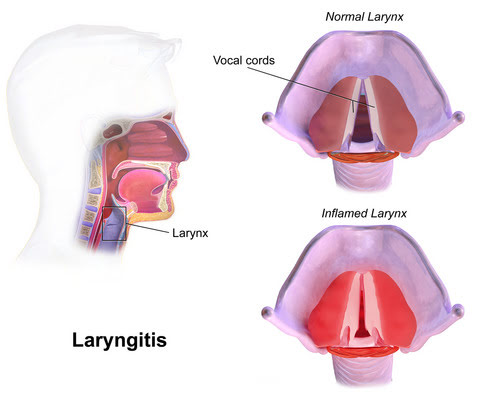
Laryngitis: Key Facts at a Glance
Laryngitis is inflammation of the voice box (larynx) and the vocal folds inside it. The swelling disrupts the normal vibration of the vocal cords, causing hoarseness, a lower-pitched voice, or a complete loss of voice. Most cases follow a cold or flu virus and get better on their own. Laryngitis itself is not contagious, but the virus behind it can spread to others.
- Acute or chronic: Acute laryngitis usually clears up within three to seven days, although mild hoarseness can linger for up to two weeks while the tissues heal. Laryngitis that lasts longer than three weeks is called chronic and is usually driven by an ongoing irritant, such as tobacco smoke, acid reflux, or continued strain on the voice.
- Main symptoms: A raspy, breathy, strained, or muffled voice, a lower pitch, or a complete loss of voice; a tickling, raw, or dry throat; a sore throat; the feeling of a lump or trapped mucus in the throat; and a dry, hacking cough. Talking usually makes the voice worse.
- Common causes: Most acute cases come from viruses that cause colds and flu, such as rhinoviruses, adenoviruses, influenza, and parainfluenza viruses. Other causes include shouting, cheering, or forceful singing; cigarette smoke, vaping, and chemical fumes; and stomach acid reaching the throat (laryngopharyngeal reflux), often without heartburn. Bacterial infection is rare and usually follows a viral one.
- Home care: Rest the voice and avoid shouting. Do not whisper, because whispering strains the vocal cords; if you must talk, speak gently at a normal pitch. Drink plenty of water, breathe cool, moist air from a humidifier, and avoid caffeine and alcohol, which dry the throat. Take a sip of water or swallow instead of clearing the throat.
- Medicines: Antibiotics do not help viral laryngitis and do not speed recovery; they are reserved for confirmed bacterial infections. Oral steroids are not used routinely because of their side effects. When reflux is confirmed as the cause, treatment starts with lifestyle changes, such as not eating for at least three hours before bed.
- When to get help: A young child’s airway is much narrower, so swelling can quickly make breathing hard. Seek emergency care right away for noisy, high-pitched breathing (stridor) while resting, the skin pulling in around the ribs or neck with each breath, drooling or trouble swallowing saliva, or sitting upright and leaning forward to breathe. See an ear, nose, and throat specialist if hoarseness lasts longer than three weeks.
- Prevention: Wash hands regularly with soap and water, stay up to date on flu and other recommended vaccines, avoid cigarette smoke and secondhand smoke, use a microphone instead of straining the voice in noisy rooms, and run a cool-mist humidifier in dry weather.
Laryngitis: Key Causes, Common Symptoms, and Evidence-Based Treatments
The human vocal mechanism is an extraordinarily refined biological instrument that relies on the coordinated interaction of respiratory airflow, intrinsic laryngeal muscles, and delicate mucosal folds. When acute inflammation, localized infection, or persistent mechanical irritation disrupts this system, the resulting clinical condition is known as Laryngitis. This upper airway pathology can alter acoustic stability within hours, reducing a clear speaking voice to an unsteady, breathy rasp or producing complete vocal silence. While acute laryngeal swelling is often considered an uncomfortable, self-limiting consequence of a common winter viral cold or an evening of cheering at a crowded sporting event, inflammatory processes of the voice box span a broad clinical spectrum. A nuanced medical understanding of laryngeal microanatomy, phonatory biomechanics, differential diagnostic criteria, and evidence-based therapeutic protocols is vital for achieving rapid vocal recovery while avoiding chronic structural injury.
Clinical Care Card: Triage and First-Line Management
Acute vocal changes presenting for fewer than seven days are overwhelmingly benign, self-limiting, and viral in origin. First-line management demands immediate, complete vocal rest, aggressive systemic hydration, ambient steam humidification, and the strict avoidance of whispering or dry throat clearing. Routine empirical antibiotic prescriptions are clinically inappropriate because antibacterial agents provide no therapeutic benefit against viral pathogens and do not accelerate recovery. Any patient presenting with resting inspiratory stridor, tachypnea, severe odynophagia, inability to manage oral secretions, or dysphonia persisting beyond three weeks requires prompt fiberoptic visualization to exclude airway compromise, invasive bacterial infection, or underlying structural lesions.
The history of diagnosing and managing upper airway inflammation dates back to classical medical antiquity. Hippocrates documented the earliest clinical descriptions of fatal upper airway obstruction nearly twenty-four centuries ago, observing patients with sudden high fever, painful swallowing, jaw swelling, and rapid suffocation when attempting to lie flat. Centuries later, in 1791, physicians published the first systematic description of acute supraglottic inflammation, and in 1799, the first President of the United States, George Washington, died following a fulminant throat infection that retrospective epidemiological analyses attribute to acute epiglottitis. The invention of the mirror laryngoscope in the 1850s marked a major clinical milestone by allowing physicians to inspect the vocal cords and hypopharynx under direct illumination. Over the course of the twentieth century, microbiological discoveries and widespread conjugate vaccination campaigns reduced the incidence of invasive pediatric airway infections. Nevertheless, laryngeal inflammation remains one of the most common respiratory diagnoses encountered across emergency departments, general practices, and specialized laryngology clinics worldwide.
| Clinical Parameter | Acute Laryngeal Inflammation | Chronic Laryngeal Inflammation |
| Typical Duration | Lasts fewer than 3 weeks; often resolves in 3 to 7 days | Persists continuously or intermittently beyond 3 weeks |
| Primary Pathophysiology | Reversible interstitial edema and neutrophilic infiltration | Mononuclear infiltration, squamous metaplasia, tissue remodeling |
| Predominant Etiology | Upper respiratory viral infection or acute vocal abuse | Ongoing acid reflux, tobacco smoke, persistent phonotrauma |
| Acoustic Vocal Impact | Sudden hoarseness, pitch depression, aphonia | Chronic vocal raspiness, reduced dynamic range, vocal fatigue |
| Endoscopic Findings | Diffuse mucosal erythema and bilateral vocal fold swelling | Nodules, contact granulomas, mucosal thickening, pachydermia |
| Standard Treatment | Absolute voice rest, systemic fluids, cool humidification | Trigger elimination, speech pathology therapy, targeted medication |
Anatomical Foundations and Pathophysiology of Laryngitis
To recognize how inflammatory cascades compromise voice production, one must evaluate the functional anatomy of the larynx. Positioned at the superior entrance of the trachea, the larynx serves as a vital gatekeeper for the human respiratory tree. It prevents aspiration of food and fluids into the lower airways during swallowing, regulates intrathoracic pressures during heavy physical exertion, and houses the vocal folds necessary for oral communication. Suspended within this cartilaginous framework are the bilateral true vocal cords (vocal folds). These specialized structures feature a layered microarchitecture consisting of a muscular base (the thyroarytenoid muscle), an extracellular matrix known as the lamina propria, and an outer covering of stratified squamous epithelium.
During resting respiration, the vocal folds abduct widely to maintain an open glottic airway. During speech, intrinsic laryngeal adductor muscles draw the vocal folds together along the midline. As pressurized air from the lungs moves upward through this narrowed glottic space, it initiates rapid, self-sustaining mucosal wave vibrations driven by subglottic air pressure and the aerodynamic Bernoulli effect. These rapid vibratory cycles generate fundamental acoustic frequencies that are subsequently shaped into distinct consonants and vowels by the tongue, palate, pharyngeal walls, and lips.
When an inflammatory response occurs, this delicate biomechanical balance fails. Localized capillary dilation, endothelial permeability, and cellular exudation promote tissue swelling across the superficial layer of the lamina propria, an anatomical zone known as Reinke’s space. The accumulation of fluid increases both the mass and the stiffness of the vibrating margins of the vocal folds. As a result, the cords can no longer approximate smoothly or sustain symmetric mucosal wave oscillations. Pressurized air leaks through the incompletely closed glottic chink, generating acoustic turbulence, pitch perturbation (jitter), amplitude instability (shimmer), and the noticeable auditory roughness recognized as dysphonia. When physicians evaluate this broad laryngitis disease process, they divide cases into acute and chronic forms based on clinical duration and histopathological characteristics.
Cellular Mechanisms in Acute Laryngitis
Acute inflammation of the vocal cords is defined by a rapid onset of symptoms that run a self-limiting course, resolving within three weeks. In everyday practice, uncomplicated acute episodes resolve within three to seven days. The underlying histopathology is characterized by acute microvascular congestion and neutrophilic migration into the mucosal stroma of the vocal fold cover. Inflammatory cytokines cause localized vasodilation, resulting in macroscopic mucosal redness and interstitial edema.
Because the connective tissue of Reinke’s space is pliable, fluid accumulates rapidly between the basement membrane and the underlying vocalis muscle. This physical swelling dampens vocal cord oscillation, lowering the baseline pitch of the speaking voice. Because cellular architecture remains intact without irreversible structural remodeling, resting the strained voice box and clearing the causative pathogen allow interstitial fluid to drain through the laryngeal lymphatics, restoring normal tissue pliability.
Histopathological Shifts in Chronic Laryngitis
When laryngeal inflammation persists for longer than three weeks, the clinical presentation is classified as chronic. Chronic laryngeal irritation involves cellular changes, matrix remodeling, and persistent mononuclear infiltration dominated by lymphocytes, plasma cells, and fibroblasts. Sustained physical stress from speaking over swollen cords or chemical exposure from gastric juices and cigarette smoke triggers adaptive structural changes throughout the laryngeal mucosa. The normal pseudostratified ciliated columnar epithelium of the respiratory tract can undergo squamous metaplasia, creating irregular, thickened mucosal surfaces.
Over time, active fibroblasts deposit dense collagen fibers, altering the delicate balance of elastin and hyaluronan within Reinke’s space. This chronic remodeling stiffens the oscillating edge of the vocal fold, creating vocal strain, loss of upper pitch registers, and phonatory fatigue. Chronic inflammation can also encourage localized benign structural lesions, such as fibrotic vocal nodules, gelatinous polyps, contact ulcers, or generalized polypoid degeneration (Reinke’s edema).
Glottic Edema and Distorted Mucosal Wave Dynamics
Fluid accumulation within the vocal fold cover increases tissue inertia, meaning the folds require significantly higher subglottic air pressure from the lungs to initiate and sustain oscillation. As a patient strains to overcome this physical resistance, intrinsic muscle tension rises sharply, creating throat strain, physical fatigue, and neck discomfort during speech.
Squamous Metaplasia and Ciliary Mucus Stasis
When the delicate laryngeal mucosa is subjected to chronic chemical irritants like tobacco smoke or aerosolized digestive enzymes, its protective ciliary clearance mechanism fails. Without functional cilia, thick mucus adheres directly to the vocal cords. This sticky accumulation triggers an involuntary, repeated urge to clear the throat, which repeatedly slams the vocal folds together and intensifies tissue irritation.
Symptoms and Clinical Presentation of Laryngitis
The clinical presentation of vocal cord inflammation involves both audible acoustic changes and localized anterior neck discomfort. Because voice production relies on balanced bilateral vocal fold vibrations, even minor swelling along the mucosal margins produces recognizable changes in vocal timbre, volume, and resonance. Patients experience an array of distinct laryngitis symptoms that vary based on the underlying cause, the patient’s vocal demands, and the anatomical structures involved.
Primary Phonic and Sensory Manifestations of Laryngitis
The primary clinical hallmark of laryngeal swelling is dysphonia, an auditory change in voice quality. The voice often sounds raspy, breathy, strained, or muffled. When vocal fold swelling reaches advanced stages, the mucosal edges become so immobile that aerodynamic vibration ceases entirely, leaving the patient completely aphonic and capable of producing only unvoiced whispers.
Beyond audible vocal deterioration, patients frequently report uncomfortable sensory symptoms localized to the larynx and hypopharynx. Because sensory branches of the superior and recurrent laryngeal nerves innervate these tissues, inflammation creates sensations of throat tickling, rawness, dryness, and a continuous globus sensation—the perception of a persistent lump or trapped mucus in the throat. Patients frequently report a painful sore throat and loss of voice that worsens with continued speaking. A dry, hacking, non-productive cough is common, provoked by subepithelial cough receptors exposed to environmental air currents.
Critical Warning Indicators and Emergency Airway Risks
While uncomplicated vocal inflammation in adults is an uncomfortable yet self-limiting disorder, laryngeal swelling in pediatric patients demands careful clinical oversight. The infant and pediatric airway is structurally narrower, funnel-shaped, and positioned higher in the neck. The subglottic space inside the cricoid cartilage forms the narrowest portion of the young child’s airway and possesses a complete, unyielding cartilaginous ring. Consequently, even one millimeter of inflammatory mucosal edema produces a severe reduction in airway cross-sectional caliber, dramatically increasing airflow resistance.
| Airway Feature | Pediatric Airway Dynamics | Adult Airway Dynamics |
| Narrowest Anatomic Point | Subglottis at the rigid cricoid ring | Glottic chink between true vocal folds |
| Laryngeal Cartilage Shape | Funnel-shaped, pliable, anteriorly inclined | Cylindrical, rigid, vertically oriented |
| Mucosal Swelling Impact | 1 mm edema causes a 50% luminal reduction | 1 mm edema causes minimal airway resistance change |
| Primary Airway Risk | Rapid stridor, barking cough, acute distress | Isolated hoarseness, aphonia, throat discomfort |
| Common Clinical Entities | Viral croup, subglottitis, epiglottitis | Acute viral corditis, reflux laryngitis, phonotrauma |
| Emergency Triage Priority | High risk for rapid respiratory decompensation | Low obstruction risk unless supraglottitis occurs |
Clinical Warning: Pediatric Airway Obstruction
Laryngeal swelling in young infants can escalate rapidly from mild vocal huskiness to dangerous respiratory exhaustion. Parents and clinicians must seek emergency care immediately upon recognizing the following clinical signs:
- Inspiratory Stridor: A harsh, high-pitched musical sound heard during inhalation, indicating critical narrowing of the glottic or subglottic lumen.
- Seal-Like Barking Cough: A resonant, deep cough caused by subglottic mucosal edema vibrating within the cricoid ring.
- Chest Wall Retractions: Marked inward sinking of the suprasternal notch, intercostal spaces, and substernal abdomen during inhalation.
- Excessive Drooling and Odynophagia: Inability or refusal to swallow oral secretions due to severe supraglottic throat pain, pointing to acute epiglottitis.
- Tripod or Sniffing Posture: Instinctively sitting upright, leaning forward with the neck hyperextended, to maintain airway patency.
Clinical Progression from Sore Throat to Lost Voice
The progression from a mild pharyngeal ache to complete vocal loss follows a predictable trajectory in viral upper respiratory infections. Infection initially colonizes the mucosal surfaces of the nasopharynx and tonsillar pillars, producing localized pharyngitis, mild rhinorrhea, and odynophagia. Over the subsequent forty-eight hours, descending viral shedding and mucosal secretions travel down the posterior pharyngeal wall into the laryngeal vestibule. Once the true vocal cords become infected, inflammation quickly causes vocal instability, pitch alterations, and eventual loss of phonation.
Inspiratory Stridor and Glottic Narrowing
Stridor represents turbulent airflow rushing through a narrowed airway passage. When inflammation is confined to the true vocal cords, resting stridor is rare because the glottic aperture remains sufficiently wide for tidal breathing. However, if edema spreads to the subglottic space—as observed in acute laryngotracheobronchitis—or affects the supraglottic tissues, cross-sectional airway diameter drops precipitously. The presence of resting stridor indicates significant luminal narrowing and requires prompt medical evaluation.
Causes and Contributing Triggers of Laryngitis
A clinical analysis of underlying laryngitis causes reveals a wide variety of infectious pathogens, environmental exposures, and mechanical strain. A precise etiological diagnosis is necessary to select appropriate therapies and avoid ineffective treatments.
| Etiological Class | Specific Causative Triggers | Primary Clinical Features |
| Upper Airway Viruses | Adenoviruses, Influenza A/B, Parainfluenza, Rhinoviruses | Acute hoarseness, coryza, low fever, self-limiting |
| Secondary Bacteria | S. pneumoniae, Group A Streptococcus, M. catarrhalis | Purulent sputum, high fever, secondary to viral cold |
| Fungal Pathogens | Candida albicans, Aspergillus species | White mucosal plaques, burning throat, common with steroid inhalers |
| Aerosol Irritants | Cigarette smoke, vaping, chemical solvents, dust | Chronic mucosal thickening, loss of vocal range, dryness |
| Acute Phonotrauma | Shouting, screaming, forceful singing without training | Sudden aphonia, vocal fold micro-hematoma, localized swelling |
| Gastric Micro-Reflux | Retrograde acid and pepsin exposure past the esophagus | Morning hoarseness, globus sensation, chronic throat clearing |
Viral Pathogens Inducing Viral Laryngitis
The vast majority of acute infectious cases represent viral laryngitis occurring within the context of a generalized upper respiratory tract infection. Respiratory viruses enter the host through aerosolized droplet inhalation or direct hand-to-mucosa inoculation. These pathogens target and infect the epithelial cells of the respiratory mucosa, replicating intracellularly and triggering host immune responses that cause cell death, mucosal shedding, and inflammation.
- Adenoviruses: Frequently cause acute pharyngolaryngeal inflammation, often characterized by follicular pharyngitis, persistent high fever, hoarseness, and anterior cervical lymphadenopathy.
- Influenza A and B: Highly aggressive pathogens that induce extensive epithelial desquamation throughout the respiratory tree. Influenza infections cause severe laryngeal erythema, high fever, systemic myalgias, and carry an elevated risk for secondary bacterial pneumonia or bacterial tracheitis.
- Parainfluenza Viruses (Types 1, 2, and 3): The predominant viral drivers of childhood croup (laryngotracheobronchitis), with type 1 serving as the primary cause of biennial autumn epidemics. Parainfluenza targets the subglottic mucosa, provoking extensive edema within the non-distensible cricoid ring.
- Rhinoviruses and Coronaviruses: The primary drivers of the common cold, these agents routinely trigger mild, transient laryngeal hyperemia, mild vocal huskiness, sneezing, and watery rhinorrhea.
- Respiratory Syncytial Virus (RSV): While noted for inducing lower respiratory bronchiolitis in infants, RSV also causes upper airway inflammation across all age brackets.
Primary bacterial infection of the vocal cords is exceptionally rare in modern clinical practice. Instead, bacterial involvement almost universally arises as a secondary superinfection following viral damage to the laryngeal mucosa. Organisms implicated in secondary bacterial laryngeal infections include Streptococcus pneumoniae, Group A beta-hemolytic Streptococcus, Moraxella catarrhalis, and non-typeable Haemophilus influenzae. Fungal laryngeal infections, predominantly caused by Candida albicans, occur almost exclusively in immunocompromised patients or individuals using inhaled corticosteroids for asthma management without using a spacer or performing post-inhalation mouth rinsing.
Environmental Irritants and Mechanical Phonotrauma
Non-infectious etiologies account for a substantial percentage of voice disorders and are the leading drivers of chronic vocal disability. Mechanical vocal trauma represents the most common non-microbial insult. Excessive shouting, cheering, sustained speaking over ambient background noise, or forceful singing without adequate breath support can slam the vocal folds together with extreme collision forces. This acute mechanical trauma causes focal subepithelial capillary rupture, microvascular hemorrhage, and localized mucosal edema.
Environmental and chemical exposures also contribute significantly to chronic laryngeal irritation:
- Combustion Smoke and Secondhand Vapors: Cigarette, cigar, and cannabis smoke contain thousands of particulate toxins and thermal combustion products that dry and burn the vocal folds. Chronic exposure damages ciliary clearance, thickens the epithelium, and causes the deep, raspy voice historically termed smoker’s laryngitis.
- Industrial Chemical Fumes: Occupational exposure to volatile organic solvents, ammonia, acid fumes, paints, and agricultural dust causes chronic chemical irritation and mucosal inflammation.
- Alcohol Overuse: Heavy ethanol intake dilates mucosal blood vessels and promotes systemic dehydration, depleting the thin serous fluid layer that cushions the vocal cords during phonation.
- Recurrent Respiratory Papillomatosis (RRP): Caused by mucosal infection with Human Papillomavirus (most commonly HPV types 6 and 11), RRP produces benign, cauliflower-like epithelial growths on the vocal folds. These lesions disrupt glottic closure and cause progressive hoarseness, stridor, and respiratory distress that frequently require repeated surgical intervention.
Epithelial Damage and Secondary Bacterial Invasion
When viral pathogens destroy the ciliated columnar cells of the respiratory tract, they break down the primary defense barrier of the airway. As host cells die, the exposed basement membrane becomes susceptible to colonizing bacteria from the pharynx. In individuals weakened by exhaustion or viral illness, common bacteria such as Streptococcus pneumoniae or Moraxella catarrhalis can invade the damaged tissue, turning a mild viral inflammation into a purulent bacterial infection.
Phonotrauma and Mucosal Fluid Loss
Healthy vocal cord oscillation relies on a thin, protective fluid barrier that lubricates the delicate mucosal surfaces as they make contact. Inhaling smoke, airborne dust, or dry heated air rapidly strips away this protective moisture. Without adequate fluid lubrication, mechanical friction increases dramatically during speech, shearing the delicate surface cells of Reinke’s space and triggering an inflammatory response even during routine talking.
Acid Reflux and Gastroesophageal Factors in Laryngitis
A major non-infectious driver of chronic voice box irritation is the retrograde ascent of gastric acid and digestive enzymes past the upper esophageal sphincter, a condition designated as Laryngopharyngeal Reflux (LPR) or reflux laryngeal inflammation. Unlike classic gastroesophageal reflux disease (GERD)—which is characterized by frequent lower esophageal acid exposure and daytime heartburn—LPR frequently manifests as silent reflux, lacking retrosternal burning entirely.
When acidic gastric juices and pepsin enter the hypopharynx, they directly contact laryngeal tissues that lack the protective bicarbonate coating and clearing mechanisms native to the distal esophagus. Pepsin binds to laryngeal epithelial cells, where it remains dormant until reactivated by acidic exposures, triggering local tissue damage, cellular inflammation, and vocal fold edema.
Clinical Profile: LPR Versus Classic GERD
While both conditions stem from gastric contents escaping the stomach, their presentations differ fundamentally:
- Primary Symptom Profile: GERD presents with retrosternal pyrosis (heartburn) and acid regurgitation; LPR presents with throat clearing, hoarseness, globus sensation, and chronic cough without heartburn.
- Postural Timing of Episodes: GERD occurs predominantly in the supine position during sleep; LPR episodes occur predominantly in the upright position during daytime activity.
- Epithelial Susceptibility: The esophagus can withstand dozens of daily reflux events without structural damage; the delicate laryngeal epithelium develops inflammation after just a few acid-pepsin exposures per week.
- Therapeutic Response: GERD symptoms respond predictably to standard-dose acid suppression; LPR symptoms respond inconsistently to empirical proton pump inhibitors in randomized trials.
Diagnostic Scoring Systems and Controversies in Reflux Laryngitis
In everyday outpatient otolaryngology, physicians often rely on clinical scoring systems to diagnose reflux-mediated vocal inflammation: the patient-reported Reflux Symptom Index (RSI) and the clinician-rated Reflux Finding Score (RFS). An RSI score exceeding 13 points (derived from self-ratings of hoarseness, throat clearing, globus sensation, chronic cough, and excess mucus) is traditionally deemed indicative of significant reflux. Concurrently, an RFS above 7 points—scored during fiberoptic laryngoscopy based on subglottic edema (pseudosulcus vocalis), ventricular obliteration, erythema, vocal cord edema, and posterior commissure hypertrophy—is widely used to confirm reflux laryngeal disease.
However, systematic reviews analyzing 24-hour double-probe (pharyngeal and esophageal) pH monitoring have revealed major diagnostic discrepancies. Objective measurements indicate that documented pharyngeal reflux (PR) events occur in roughly 22.9 percent of healthy, asymptomatic individuals, while occurring in only 38.3 percent of patients clinically diagnosed with reflux laryngeal inflammation. This small numerical difference fails to achieve statistical significance (P = 0.079), underscoring that double-probe pH monitoring cannot reliably differentiate symptomatic patients from normal controls. Furthermore, large-scale, placebo-controlled randomized clinical trials have repeatedly failed to demonstrate a clear therapeutic superiority of proton pump inhibitors (PPIs) over placebo in resolving persistent laryngeal symptoms. This clinical mismatch suggests that reflux-induced vocal swelling is frequently overdiagnosed in routine clinical settings.
| Diagnostic Tool | Clinical Evaluation Target | Documented Scientific Limitations |
| Reflux Symptom Index (RSI) | Self-reported survey assessing 9 throat-specific symptoms | High sensitivity but low specificity; any throat irritant elevates the score above 13 |
| Reflux Finding Score (RFS) | Endoscopic grading of laryngeal erythema and posterior edema | Significant observer bias; identical signs appear in up to 86% of healthy individuals |
| Pseudosulcus Identification | Inspecting for subglottic mucosal edema below vocal cords | Visible in up to 49% of healthy volunteers; not specific to acid reflux |
| Dual-Probe 24-Hour pH | Recording pharyngeal acid events above the upper sphincter | Detects events in 38.3% of suspected patients vs. 22.9% of healthy controls (P = 0.079) |
| Empirical High-Dose PPIs | Twice-daily acid suppression prescribed for 8 to 12 weeks | Randomized trials show no superiority over placebo; risks drug dependency |
Diagnostic Specificity Deficits of the Reflux Finding Score
A critical limitation of the Reflux Finding Score is that posterior laryngeal erythema and interarytenoid pachydermia are non-specific signs. Environmental dust, seasonal allergies, persistent mouth breathing, chronic vocal strain, and normal anatomic variations regularly produce identical mucosal changes. Relying solely on visual inspection during laryngoscopy frequently results in misattributing unrelated vocal disorders to acid reflux.
Therapeutic Outcomes of Empirical Proton Pump Inhibitors
The routine prescription of empirical, high-dose proton pump inhibitors for unverified reflux laryngeal inflammation carries notable medical risks. Prolonged hypochlorhydria impairs enteral absorption of calcium, magnesium, and vitamin B12, increases susceptibility to enteric infections like Clostridioides difficile, and creates severe rebound acid hypersecretion upon sudden drug withdrawal. Acid suppression therapy should remain targeted and continuously reevaluated.
Differential Diagnosis and Emergency Airway Distinctions in Laryngitis
When patients present with dysphonia, throat discomfort, and cough, primary care physicians and otolaryngologists must differentiate uncomplicated vocal inflammation from life-threatening upper airway infections and progressive structural pathology.
| Condition | Primary Site of Involvement | Defining Clinical Triad | Typical Patient Demographics | Level of Airway Danger |
| Laryngitis | True vocal folds (glottis) | Dysphonia, throat clearing, raw throat | All age groups, post-viral or vocal strain | Negligible in isolated adult cases |
| Viral Croup (LTB) | Subglottic larynx and trachea | Seal-bark cough, stridor, chest retractions | Children aged 6 months to 3 years | Moderate; responsive to steroids |
| Acute Epiglottitis | Supraglottic structures | Drooling, dysphagia, tripod posture | Adults (mean age 40s) & unvaccinated kids | Extreme emergency; rapid fatal closure |
| Bacterial Tracheitis | Tracheal subglottic mucosa | High toxic fever, purulence, failed croup care | Children with prior respiratory viruses | High; frequently requires intubation |
| Laryngeal Carcinoma | Glottic/supraglottic tissue | Progressive hoarseness, weight loss, hemoptysis | Adults with tobacco/alcohol exposure | Progressive mechanical airway compromise |
Comparing Supraglottitis, Croup, and Tracheitis with Laryngitis
Because inflammation can arise across various anatomical compartments of the airway, differential diagnosis must systematically consider epiglottitis, croup, and bacterial tracheitis:
- Acute Epiglottitis (Supraglottitis): A rapidly progressive, life-threatening bacterial infection of the epiglottis, aryepiglottic folds, and surrounding supraglottic tissues. While historically an infection of young children caused by Haemophilus influenzae type b (Hib), widespread vaccination has made epiglottitis predominantly an adult disease caused by Streptococcus pneumoniae, Group A Streptococcus, and Staphylococcus aureus. Adults present with severe sore throat out of proportion to physical findings, odynophagia, inability to swallow secretions, high fever, and a muffled “hot potato” voice. Unlike patients with vocal fold inflammation who have a raspy voice and breathe easily, epiglottitis patients appear toxic and can experience rapid, fatal airway closure.
- Viral Croup (Acute Laryngotracheobronchitis): The primary cause of acute upper airway obstruction in children aged 6 months to 3 years. Caused predominantly by parainfluenza virus type 1, croup involves inflammatory edema in the subglottic mucosa directly beneath the vocal folds. It presents with a classic seal-bark cough, inspiratory stridor, and chest wall retractions that worsen at night. Anteroposterior neck radiographs reveal subglottic tapering known as the “steeple sign”.
- Bacterial Tracheitis: A rare but dangerous invasive bacterial infection of the subglottic tracheal mucosa, frequently developing as a secondary superinfection following viral influenza or croup. Children present with high fevers, a toxic appearance, thick mucopurulent secretions, and progressive airway obstruction that fails to respond to nebulized racemic epinephrine and corticosteroids. Direct visualization in an operating room is required to secure the airway and clear purulent tracheal pseudomembranes.
Malignancy and Structural Vocal Fold Lesions Beyond Laryngitis
When vocal impairment persists for more than three weeks, physicians must look beyond simple mucosal inflammation. Chronic hoarseness is the most consistent initial warning sign of laryngeal cancer, predominantly squamous cell carcinoma of the vocal folds. Patients with significant histories of tobacco use, heavy alcohol intake, or unexplained weight loss require immediate referral to an otolaryngologist for transnasal flexible laryngoscopy or videostroboscopy.
Direct optical visualization allows early detection of vocal fold leukoplakia, erythroplasia, exophytic fungating masses, and vocal cord fixation. Treating persistent dysphonia with repeated empirical courses of antibiotics or anti-reflux medications without visual inspection risks delaying an early oncologic diagnosis when conservative, voice-preserving therapy is most successful.
Subglottic Edema and Narrowing in Pediatric Airways
Airflow dynamics dictate that airway resistance changes inversely with the fourth power of the internal radius. In small infants, a single millimeter of inflammatory swelling within the narrow subglottic cricoid ring reduces total airway area by half and increases the effort of breathing sixteen-fold. This anatomical reality explains why a mild viral infection that produces simple voice huskiness in an adult can cause severe respiratory distress and stridor in a young toddler.
Videostroboscopy and Mucosal Wave Assessment
Standard continuous light laryngoscopy provides clear images of vocal fold color, swelling, and macroscopic growths, but it cannot evaluate the microscopic movement of the vocal cover during phonation. Videostroboscopy synchronizes a rapid strobe flash with the fundamental frequency of the patient’s voice, producing a simulated slow-motion view of the mucosal wave. This allows the clinician to identify focal stiffening, early invasive carcinomas, intracordal cysts, or subtle vocal nodules that cannot be detected with regular light.
Evidence-Based Management and Laryngitis Treatment
Managing vocal cord inflammation requires a structured approach centered on non-pharmacological care, avoidance of inappropriate medications, and targeted therapies for identified underlying conditions. The primary objective of standard laryngitis treatment is to alleviate mucosal irritation, facilitate tissue healing within the lamina propria, and restore vocal function. Modern clinical guidelines emphasize that conservative laryngitis treatments outperform aggressive pharmacotherapy in uncomplicated presentations.
Voice Health Protocol: Essential Home Practices
- Enforce Absolute Voice Rest: Refrain from unnecessary talking, shouting, or singing for 48 to 72 hours. Continued voice use across swollen vocal folds delays tissue healing and increases the risk of vocal cord hemorrhage.
- Never Whisper to Communicate: Whispering forces the front of the vocal folds tightly together while leaving the back open, concentrating mechanical friction along the margins and worsening swelling. If you must talk, speak gently at a normal pitch.
- Maintain Systemic Hydration: Drink plenty of water throughout the day to keep mucosal secretions thin and easily cleared. Breathing in cool mist or taking warm steam showers delivers direct moisture to raw airway tissues.
- Eliminate Mucosal Drying Agents: Avoid caffeinated beverages, alcohol, and over-the-counter decongestants. These substances dry out airway tissues and increase vocal cord friction during speech.
Conservative Care and Behavioral Regimens
The cornerstone of conservative treatment for laryngitis is resting the voice. When the vocal cords are swollen, every vocal fold collision causes mechanical shearing across fragile epithelial layers. Strict voice rest eliminates this physical friction, allowing lymphatic channels to drain interstitial edema and preventing microvascular hemorrhage in Reinke’s space.
Systemic hydration is equally essential. Adequate oral fluid intake thins the protective mucus blanket covering the respiratory tract, allowing it to coat the colliding cords with every phonatory cycle. Direct surface hydration through steam inhalation or personal cool-mist humidifiers provides symptomatic relief by soothing irritated mucosal surfaces.
| Phase of Care | Clinical Action Plan | Practical Management Recommendations |
| Acute Phase (Days 1 to 7) | Complete vocal rest and hydration | Silence phone calls, stop shouting, run cool-mist humidifiers, avoid whispering |
| Subacute Phase (Days 8 to 21) | Controlled, gentle vocal re-entry | Reintroduce speaking at low volume, maintain 2–3 liters daily fluid, stop throat clearing |
| Chronic Phase (Beyond 21 Days) | Formal laryngological evaluation | Flexible fiberoptic endoscopy, videostroboscopy, speech pathology therapy |
| Refractory Phase | Targeted intervention and microsurgery | Phonomicrosurgery for mature polyps, medical antireflux care, allergen avoidance |
Judicious Medical Interventions
Pharmacotherapy plays an adjunct role in managing acute laryngeal conditions and must be applied judiciously based on clinical evidence:
- Corticosteroids: A brief course of oral corticosteroids (such as prednisone, methylprednisolone, or dexamethasone) can rapidly reduce laryngeal hyperemia and vocal fold edema. However, systemic steroids carry distinct adverse effects (hyperglycemia, psychiatric disturbances, gastrointestinal ulceration) and should not be used routinely. They are clinically reserved for professional voice users (actors, singers, speakers) facing unavoidable performance commitments, or individuals exhibiting acute airway compromise. Notably, research indicates that inhaled budesonide achieves laryngeal anti-inflammatory outcomes comparable to oral corticosteroids while minimizing systemic drug exposure.
- Antibiotics: In uncomplicated acute vocal cord inflammation, routine antibiotic administration is clinically inappropriate. Multiple randomized, double-blind, placebo-controlled trials and Cochrane systematic reviews confirm that antibiotics fail to accelerate symptom resolution or shorten dysphonia duration in viral laryngeal infections. Their inappropriate use drives antimicrobial resistance, escalates healthcare expenditures, and exposes patients to adverse drug reactions. Antibiotic therapy is reserved strictly for laboratory-confirmed bacterial infections or cases complicated by purulent tracheobronchial superinfections.
- Antireflux Medications and Diet: For patients diagnosed with confirmed LPR, therapeutic management begins with lifestyle adjustments: elevating the head of the bed by 15 centimeters, stopping food intake at least three hours before retiring to bed, and eliminating dietary triggers (chocolate, peppermint, citrus, tomatoes, caffeine, and fatty meals). If lifestyle measures prove insufficient, a targeted course of proton pump inhibitors or H2-receptor antagonists may be utilized.
- Voice Therapy and Surgical Management: For chronic phonotrauma, collaborating with a licensed speech-language pathologist provides behavioral voice therapy. Patients learn resonant voice techniques, vocal cord relaxation exercises, and optimal breath support to eliminate phonatory strain. When persistent inflammation leads to irreversible structural lesions—such as mature fibrous polyps, extensive cysts, or severe Reinke’s edema refractory to medical therapy—microlaryngeal phonomicrosurgery using cold micro-instruments or pulsed lasers is employed to excise diseased tissue while preserving the layered architecture of the lamina propria.
Absolute Voice Rest and the Risk of Whispering
When experiencing acute voice loss, patients frequently whisper under the misconception that it spares the vocal cords from stress. Laryngoscopic studies reveal that whispering pushes the arytenoid cartilages tightly together while leaving the posterior glottis open. This asymmetrical posture forces high-velocity airflow across dry, swollen tissues, increasing mechanical friction and delaying recovery. Patients should maintain complete vocal silence or speak gently in a natural conversational voice.
Inappropriate Antibiotic Use and Corticosteroid Risks
The common practice of prescribing antibiotics for sudden hoarseness remains a major clinical challenge. Systematic reviews show that antibacterial drugs provide no measurable benefit for viral voice box inflammation. Unnecessary prescriptions expose patients to adverse drug effects and drive antimicrobial resistance. Similarly, corticosteroids should never be viewed as a quick fix for vocal fatigue, as masking pain without resting the voice increases the risk of vocal cord tears and permanent scarring.
Prevention and Vocal Health Guidelines for Laryngitis
Preventing laryngeal inflammation involves minimizing viral exposure, protecting the voice from biomechanical strain, and shielding the vocal tract from chemical irritants. Integrating good vocal hygiene into daily routines preserves voice quality and mitigates recurrent acute episodes.
| Preventative Strategy | Primary Mechanism of Action | Recommended Practical Implementation |
| Systemic Hydration | Reduces mucus thickness and vocal cord friction | Drink 2 to 3 liters of pure water daily |
| Hand Sanitization | Limits viral transmission to respiratory passages | Wash hands regularly with soap and water |
| Microphone Amplification | Decreases phonation collision velocity | Use a portable microphone when teaching or presenting |
| Room Humidification | Keeps airway linings moist in dry environments | Run a cool-mist humidifier in the bedroom overnight |
| Smoke Avoidance | Prevents thermal and chemical airway irritation | Quit smoking and avoid secondhand vapor exposure |
| Swallow Replacement | Prevents aggressive vocal cord collision damage | Take a sip of water instead of coughing or clearing |
Vocal Hygiene and Environmental Safeguards
Proactive vocal fold care begins with behavioral and environmental habits that protect the upper respiratory mucosa:
- Hand Hygiene and Immunization Protocols: Consistent handwashing with soap and water limits the spread of respiratory pathogens. Staying current with seasonal influenza, COVID-19, and pneumococcal vaccinations reduces the incidence of viral respiratory infections that can trigger vocal fold inflammation.
- Abstinence from Inhaled Toxins: Eliminating exposure to primary cigarette smoke, secondary aerosols, and cannabis vapors is vital for long-term vocal fold health. Smoke exposure causes ongoing epithelial desiccation and chronic tissue edema.
- Airway Humidification: Utilizing ultrasonic cool-mist humidifiers in bedrooms during dry winter months counteracts the desiccating effects of indoor heating systems, ensuring the mucosal linings remain moist overnight.
- Swallow Substitution Technique: Instead of forcefully clearing the throat when experiencing a tickle or globus sensation, patients should take a sip of water or execute a dry swallow. This action clears mucus from the vocal fold surfaces without slamming the edematous cords together.
Workplace Voice Ergonomics to Forestall Recurring Laryngitis
Professional voice users—including classroom teachers, fitness instructors, trial attorneys, and telephone agents—face high occupational vocal demands that place them at elevated risk for recurring laryngeal inflammation. Implementing ergonomic workplace strategies preserves vocal stamina and protects against phonotrauma:
- Acoustic Voice Amplification: Educators and public speakers should employ portable personal voice amplification systems rather than straining their voices to project across large, noisy rooms. Voice amplification allows individuals to speak at resting conversational volumes, reducing vocal cord collision impact.
- Structured Vocal Recovery Breaks: High-demand voice professionals should schedule brief intervals of complete vocal silence throughout the workday to allow microvascular recovery within the lamina propria.
- Optimized Breath Support: Engaging diaphragmatic and abdominal breath support reduces reliance on the intrinsic laryngeal muscles, preventing hyperfunctional muscle strain during extended speaking engagements.
Mucosal Hydration and Secretion Quality
The vibrating edges of the vocal folds need a smooth, thin layer of fluid to oscillate freely without friction. When a person is dehydrated, respiratory mucus turns viscous and gummy, increasing mechanical drag across the vocal folds and forcing the muscles to work harder during speech. Drinking sufficient water throughout the day thins respiratory secretions, protecting the cords from friction and speeding recovery from inflammation.
Acoustic Amplification and Eliminating Throat Clearing Trauma
Teachers frequently speak at volume levels above 75 decibels to be heard over background classroom noise. This sustained effort requires high subglottic air pressure and causes forceful collisions between the vocal folds. Using a small wireless microphone reduces required vocal effort, cutting vocal fold impact by half. Similarly, substituting a gentle swallow for forceful throat clearing avoids abrasive contact damage to the delicate oscillating edges of the cords.
Frequently Asked Questions
What is laryngitis?
It is an inflammatory condition affecting the larynx, commonly called the voice box, and its internal vocal folds. This swelling disrupts the smooth vibratory movement of the vocal cords as air passes over them from the lungs, leading to hoarseness, pitch lowering, or complete voice loss. The disorder can present as an acute, self-limiting condition lasting a few days or persist as a chronic clinical concern requiring specialized medical evaluation.
Is laryngitis contagious?
The vocal fold inflammation itself is not an independent contagious disease, but whether it spreads depends entirely on the underlying cause. When swelling develops secondary to upper respiratory viruses—such as the common cold, influenza, or adenoviruses—those viral pathogens are contagious and spread via respiratory droplets. Conversely, when vocal cord inflammation stems from mechanical voice strain, acid reflux, or environmental chemical exposure, it is non-infectious and cannot be transmitted to others.
Is laryngitis communicable?
The condition is not communicable across all clinical forms. Communicability is restricted to cases caused by transmissible biological agents, such as respiratory viruses or atypical bacterial pathogens. Non-infectious varieties—such as those provoked by shouting, long-term cigarette smoking, or gastroesophageal acid reflux—are non-communicable internal physical processes.
Is laryngitis spreadable?
Laryngeal swelling caused by physical vocal strain or chronic chemical irritants is not spreadable between individuals. However, when the condition arises as a symptom of a viral respiratory infection, the underlying virus spreads through aerosolized droplets, coughing, or direct contact with contaminated surfaces. Practicing frequent handwashing and staying home during active viral illnesses prevents the spread of these causative agents.
How long does laryngitis last?
Uncomplicated acute vocal fold inflammation typically resolves within three to seven days, though mild hoarseness may persist for up to two weeks as tissues heal. In contrast, chronic presentations persist for longer than three weeks, often lasting for months if underlying irritants like tobacco smoke or acid reflux remain unaddressed. Any case of hoarseness that lasts beyond three weeks requires direct visualization of the vocal cords by an ear, nose, and throat specialist.
How to treat laryngitis?
The primary step in managing vocal fold inflammation is maintaining strict voice rest, avoiding shouting, and never whispering, which strains the vocal cords. Patients should stay well-hydrated with water, inhale cool steam, run a room humidifier, and avoid drying substances like caffeine, alcohol, and decongestants. If symptoms persist or stem from chronic causes like acid reflux, a healthcare professional can tailor treatment with targeted medical therapies or referral to speech therapy.
The following posts may interest you
Infant Reflux: Causes, Symptoms, and Treatments
Colic vs Gas vs Reflux: Why Is My Baby Crying?
Effective Acid Reflux Remedies: Proven Strategies for Relief
Disclaimer: The content provided on BabyKidsHealth.com is for informational purposes only and is not intended as a substitute for professional medical advice, diagnosis, or treatment. Always seek the advice of your physician or other qualified health provider with any questions you may have regarding a medical condition.
Sources
Laryngitis (MedlinePlus, U.S. National Library of Medicine)
https://medlineplus.gov/ency/article/001385.htm
Laryngitis (NHS, UK)
https://www.nhs.uk/conditions/laryngitis/
Hoarseness (National Institute on Deafness and Other Communication Disorders, NIH)
https://www.nidcd.nih.gov/health/hoarseness
Hoarseness (Dysphonia): Healing Your Child’s Voice (American Academy of Pediatrics, HealthyChildren.org)
Croup in Young Children (American Academy of Pediatrics, HealthyChildren.org)
Epiglottitis (American Academy of Pediatrics, HealthyChildren.org)
Sore Throat Basics (Centers for Disease Control and Prevention, CDC)
https://www.cdc.gov/sore-throat/about/index.html
About Human Parainfluenza Viruses (Centers for Disease Control and Prevention, CDC)
https://www.cdc.gov/parainfluenza/about/index.html
Laryngitis, Tracheitis, Epiglottitis, and Bronchiolitis
https://link.springer.com/chapter/10.1007/978-3-319-91080-2_7
Epiglottitis, Acute Laryngitis, and Croup
https://link.springer.com/chapter/10.1007/978-3-319-74835-1_20
Reflux and Laryngitis: A Systematic Review
https://journals.sagepub.com/doi/abs/10.1016/j.otohns.2006.12.004